Advances in Animal and Veterinary Sciences
Research Article

Prevalence and Hemato-biochemical Studies on Theileriosis in Naturally Infected Cattle in Erbil Province
Khalid Aziz1*, Nazhad Qader1, Khalid Ibrahim2
1College of Agricultural Engineering Sciences, Salahaddin University-Erbil, Iraq; 2Biology department. Faculty of Science, University of Zakho, Iraq.
Abstract | This research aimed to find out the prevalence of theileriosis by Giemsa stained blood films and to deal with hemato-biochemical changes in naturally infected cattle. The overall prevalence rates were 32.75% (95/290) for Theileria infection by Giemsa stained blood smears. Clinical examination of infected cattle showed signs of fever, anorexia, congested mucous membranes, corneal opacity, emaciation, enlargement of superficial lymph nodes, pale mucous membrane, respiratory signs, yellowish diarrhea and brown coffee urine were common clinical findings. There was no significant difference associated with prevalence between gender and age (P < 0.05). The hematological parameter revealed the microcytic anemia which was associated with large reductions in RBCs (4.48±0.11) 1012/l, Hb (7.15±0.18) g/dl, PCV (17.63±0.40) %, MCV (39.69±0.58) fl, MCHC (36.98±0.67) g/l and neutrophils (3.21±0.203) %. While a significant increase in the WBCs (13.24±0.32)109/l and lymphocytes (61.42±1.84) % were found. The biochemical parameter revealed a significant increase in active serum enzyme AST (87.60± 0.12), ALT (21.70±0.07), ALP (119±0.06), and total serum bilirubin (1.13±0.01); whereas, a significant decrease in the total serum protein (6.10±0.03), albumin (2.70±0.03), calcium (7.40±0.02) and glucose (19.9±0.05) were recorded. According to the findings of this study, bovine theileriosis is a serious infectious disease in Erbil province. A more effective control program should be created to confine and manage the disease’s prevalence in the area.
Keywords | Hematological, Biochemical, Bovine Theileriosis, Erbil
Received | July 08, 2021; Accepted | October 10, 2021; Published | December 01, 2021
*Correspondence | Khalid Aziz, College of Agricultural EngineeringSciences, Salahaddin University-Erbil, Iraq; Email: khalid.aziz1@su.edu.krd
Citation | Aziz K, Qader N, Ibrahim K (2022). Prevalence and hemato-biochemical studies on theileriosis in naturally infected cattle in erbil province. Adv. Anim. Vet. Sci. 10(1): 49-54.
DOI | http://dx.doi.org/10.17582/journal.aavs/2022/10.1.49.54
ISSN (Online) | 2307-8316; ISSN (Print) | 2309-3331
Copyright © 2022 Aziz et al. This is an open access article distributed under the Creative Commons Attribution License, which permits unrestricted use, distribution, and reproduction in any medium, provided the original work is properly cited.
Introduction
Tropical theileriosis is one of the most prevalent serious problem with greatest economic and mortality impact of cattle in Iraq (Aktas et al., 2004). This disease is one of tick-borne protozoal disease caused by Theileria of the suborder Piroplasmorina which its transmitted by ticks from genus Hyalomma spp (Salih et al., 2007; Aziz and AL-Barwary, 2020). This protozoan is obligatory intracellular hemoparasites, and two species invaded to cattle T. parva (causes East Coast fever) and T. annulata (causes tropical theileriosis) (Sandhu et al., 1998; Singh et al., 2001).
The infection occurs due to the presence and multiplication of parasite inside WBCs and then RBCs and resulting in progressive and severe macrocytic hemolytic anemia (Radostits et al., 2000). The main clinical signs are enlargement of prescapular lymph nodes, fever, diarrhea, pale of mucous membrane, decrease of milk production, abortion in pregnant animals due to high fever, in appetence and lethargy (Radostits et al., 2000). Systemic alterations and lateral recumbence were seen in cows with theileriosis (Stockham et al., 2000). Whereas frequently associated with anemia, anisocytosis, pikilocytosis, and leucopenia in bovine (Ceci et al. 1997). Sharma et al. (2000) reported that the reduction of, haemoglobin (Hb), packed cell volume (PCV), differential leucocytic count (DLC) and total erythrocytic count (TEC), and this might be due to the sever intravascular haemolysis. Furthermore, Singh et al. (2001) found a significant reduction in total blood proteins, albumin, serum immunoglobulin, and albumin-to-globulin ratio in Theileria-infected calves. During the hemolytic phase in infected cattle, total serum proteins and blood glucose levels decreased (Fujinaga, 1981), while Pandey and Misra (1987) reported the protein profile normal.
Whereas changes in hemato-biochemical markers reflect the severity of theileriosis infection in cattle. The current study aimed to investigate the impact of tropical theileriosis on certain blood and serum liver function components based on the above aspects.
Material and methods
A total of 290 blood samples were collected from Coccygeal (Tail) Vein of cattle along spring season of 2021; the collected samples were from infected cattle (deferent ages, sexes and breeds) were suffered from weakness, anemia, high fever and presence of ticks. Blood samples were collected using an 18G needle into two sterile vacutainers® tubes (5 ml each), divided into two parts, one containing the anticoagulant ethylene diamine tetraacetic acid (EDTA) and the other without anticoagulant. Thin blood smears were prepared from blood samples in EDTA tubes and stained by Giemsa stain (10 %) and examined under oil immersion (X 100) according to (Chaudhri and Gupta, 2003; Aziz and Al-barwary, 2019).
Blood samples were collected in tubes with anticoagulant used for blood parameters included total red blood cell counts (TRBCs), haemoglobin concentration (Hb), packed cell volume (PCV), the mean of corpuscular volume (MCV), and corpuscular haemoglobin concentration (MCHC) and total white blood cell counts (TWBCs) by using a Haematological Analyzer (Mythic 18VET). In addition, the four field meander technique was used to determine the differential leucocytic count (DLCs) (Kelly, 1984).
While the blood samples were collected in tubes without anticoagulant used for serum separation using a centrifuge at 2500 RPM for 15 minutes and stored in -20ºC until use (Aziz and Al-Barwary., 2019). The collected sera were used for biochemical variable analysis which includes: aspartate amino transferase (AST), alanine amino transferase (ALT), alkaline phosphatase (ALP), total bilirubin (TB), total protein (TP), globulins (Glu), albumins (Alb) calcium (Ca) and glucose by using COBAS INTEGRA® 400 plus analyzer.
Statistical Analysis
All haemato-biochemical results are presented as mean + SEM and differences between groups of prevalence of theileriosis and the odds ratio and influence of risk factor (gender, age of group) were assessed using GenStat 12th Edition Software by the x 2 and Fisher’s exact tests and binomial logistic regression. A p value of <0.05 was considered significant.
Results
The overall rate of infection with bovine theileriosis by Giemsa stained blood of smears of 290 was 32.75 % (95/290). Theileria species were identified based on physical features of the merozoite in infected RBCs. Further, typical Theileria spp appeared as a small parasite measuring based on single round and double pyriform with acute or obtuse angle, and also it appeared in different morphological shapes inside the RBCs including oval, anaplasmoid, spherical, single pear and double pears shape was one the most prominent shapes as in (Figure 1).
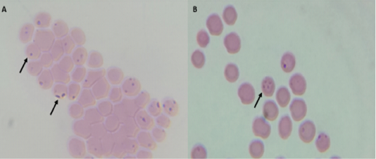
Figure 1: Morphological form of Theileria spp within RBC stained with Giemsa, examined under oil immersion lens (100X); A) Single and double pyriform, Round anaplasmoid shape; B) Maltese cross shape.
In the current research, the prevalence of Theilria spp. did not significantly differ between genders and age of cattle. Female and male recorded 17.76 % (29/63) and 11.81 % (15/127) infection rates, respectively, without significant difference P≤ 0.05. Higher rate of infection with bovine theileriosis was recorded in age group more than 1-5 years’ 21.8 % (29/133), compared with two other groups without significant differences P≤ 0.05 (Table 1).
Clinical signs
The clinically infected cattle were suffered from anorexia, dehydration, fever, hemoglobinuria, pale mucous membra-
Table 1: Sex and age-wise prevalence of Theileriosis in cattle by microscopic examination.
| Factor | No. of animals examined | No. Positive (%) | ||
| N. (%) | OR (95%CI) | p | ||
| Gender | ||||
| Female | 163 | 29 (17.78) | 1.51 (0.78-2.92) | 0.14 |
| Male | 127 | 15 (11.81) | 0.66 (0.34-1.92) | 0.14 |
| Age group | ||||
| < 1 year | 86 | 13 (15.11) | 0.81 (0.41-1.59) | 0.33 |
| 1—5 years | 133 | 29 (21.80) | 1.56 (0.86-2.83) | 0.09 |
| > 5 years | 71 | 9 (12.67) | 0.66 (0.31-1.42) | 0.19 |
| Total | 290 |
95 (32.75) |
Table 2: Clinical signs of infected cattle with theileriosis.
| Clinical signs | No. of infected cattle | % |
| Anorexia | 19 | 47.5 |
| Dehydration | 9 | 22.5 |
| Fever | 38 | 95 |
| Hemoglobinuria | 31 | 77.5 |
| Pale mucous mm. | 32 | 80 |
| Edema | 6 | 15 |
| Eye congestion and Lacrimation | 34 | 85 |
| Enlargement of prescapular lymph node | 35 | 87.5 |
| Respiratory signs | 27 | 67.5 |
| Yellowish soft diarrhea | 11 | 27.5 |
| Corneal opacity | 7 | 17.5 |
Table 3: Body temperature, respiratory rates and heart rates of infected cattle with theileriosis.
| Parameter | Control Mean± S.E | Infected Mean± S.E |
|
Body temperature/℃ |
38.9±0.140 | 40±0.081 |
| Heart rate/ min | 69.8±1.644 | 77±1.568 |
| Respiratory rate/ min | 28.2±0.377 | 37.7±0.774 |
Table 4: Hematological changes in naturally infected cattle with theileriosis compared with the control group. *indicates P≤0.05
| Parameter |
Control Mean± S.E |
Infected Mean± S.E |
|
|
RBC X 1012/l |
6.77±0.17 | 4.48±0.11* | |
| Hb g/dl | 12.43±0.40 | 7.15±0.18* | |
| PCV % | 33.30±0.61 | 17.63±0.40* | |
|
MCV fl |
45.22±0.75 | 39.69±0.58* | |
| MCHC g/l | 42.86±0.72 | 36.98±0.67* | |
|
WBC X109/l |
11.84±0.21 | 13.24±0.32* | |
| Lymphocyte % | 48.40±0.12 | 61.42±1.84* | |
| Monocyte % | 1.60±0.12 | 3.05±0.10* | |
| Neutrophil % | 28.25±2.36 | 16.85±0.77* | |
| Basophil % | 0.00±0.000 | 0.00±0.00 | |
| Eosinophil % | 2.70±0.22 |
3.22±0.20 |
|
Table 5: Biochemical changes in naturally infected cattle with theileriosis compared with the control group. *indicates P≤0.05
| Parameter |
Control Mean± S.E |
Infected Mean± S.E |
| AST U/L | 66.60±0.17 | 87.60±0.12* |
| ALT U/L | 19±0.14 | 21.70±0.07* |
| ALP U/L | 63±0.13 | 119±0.06* |
| Total bilirubin mg/dl | 0.28±0.04 | 1.13±0.01* |
| Total protein g/dl | 7.76±0.04 | 6.10 ±0.03* |
| Globulin g/dl | 4.14±0.03 | 4.1±0.02 |
| Albumin g/dl | 3.60±0.04 | 2.70±0.03* |
| Calcium g/dl | 9.80±0.07 | 7.40±0.02* |
| Glucose g/dl | 22.70±0.04 |
19.9±0.05* |
-ne, edema, eye congestion and lacrimation, enlargement of pre-scapular lymph node, respiratory signs, yellowish soft diarrhea and corneal opacity (Table 2). Besides, variations within the recoded systemic reactions were found (Table 3).
Hematological Parameter
The animals infected with bovine theileriosis were observed with microcytic hypochromic anemia, as shown in Table (4). Statistically observed a significant decrease in; red blood cells count (RBCs), mean haemoglobin amount (Hb), packet cell volume (PCV), mean corpuscular volume (MCV), mean corpuscular haemoglobin concentration (MHCH) compared to the control ones. Whereas, white blood cells count (WBCs) and lymphocyte were significantly increased.
Biochemical Parameter
Table (5) shows the biochemical parameters analysis significant increases in active serum enzyme AST, ALT, ALP and serum total bilirubin were found, while the serum total protein, albumin, calcium and glucose were significantly decreased.
Discussion
Theileriosis are considered the most important tick-born blood parasites of cattle that causes significant concern for the health and management of cattle industry especially in the tropics and subtropical area. This study supports the idea that bovine theileriosis is a major obstacle to the growth of the livestock sector in Erbil, resulting in significant economic losses due to bovine mortality and decreased production.
In the present study, the prevalence of Theileria spp. parasite in cattle in Erbil province was 32.75% which it is half percentage (59.68%) to the study in Basrah province (Minnat and Abdulwadood, 2012) and nearly double (19%) to the study in DhiQar (Abdullah and Hadi, 2021). In this study, the prevalence of Theileria spp. rates were varying between the cattle’s genders and their ages, these findings are in line with that reported by (Minnat and Abdulwadood, 2012; Khan et al., 2017). The clinical symptoms recorded in this study are identical to that reported by (Sandhu et al., 1998; Radostits et al., 2000). In addition to that, the anorexia caused by prolonging fever and lymphoid hyperplasia in the cattle at early ages infection can explain the development of superficial lymph nodes. Also, the corneal opacity of infected cattle could be due to infiltration of white blood cell (Irvin and Mawmachi, 1983).
The haematological findings of this study revealed substantial reductions in total red blood cell count, mean hemoglobin level, packed cell volume, and mean corpuscular hemoglobin concentration, all of which suggested severe anemia, which they are in line to a study by Omer et al. (2003). The findings also approve with Sandhu et al. (1998) who found normocytic normochromic anemia in calves experimentally infected with Theileria. The changes within the Leucogram might be resulted from anemia as a result of heavy infestation of blood sucking parasite (ticks) and of suppressive effect of the toxic metabolites of Theileria on the haemopiotic organs especially bone marrow hindering the leucogenesis (Durrani et al., 2008). Besides, intracellular propagation of Theileria causes excessive intravascular damage of RBCs leading to hemolytic anemia (Grewal et al., 2005; Asri & Datir 2006).
As a result of theileriosis, the biochemical parameters were altered and a substantial change within in the AST, ALT and ALP were reported which are in line with that reported previously by (Hussein et al., 2004, Ramazan, 2006). The enzymes AST and ALT are involved in the metabolism of amino acids and carbohydrates. The muscles and liver have large concentrations of these enzymes. The presence of these enzymes in the blood indicates the presence of organ necrosis (Forsyth et al., 1999). In addition, the total bilirubin levels in infected cattle increased significantly. These findings might be linked to hepatic dysfunction and, most likely, hemolytic anemia (Hussein et al., 2004). Similar finding has been achieved by others (Omer et al., 2003; Hussein et al., 2004, Ramazan, 2006).
The blood total protein and albumin levels were found to be significantly lower in the current study. These findings were similar to the previous studies (Omer et al., 2003, Hussein et al., 2004, Ramazan, 2006). Hypoproteinaemia and hypoalboumineamia are possible side effects of Theileria’s toxic metabolites, as well as liver failure. Also the observed hypoglycemia in infected cattle with theileriosis could be attributed to persistent feverish condition associated theileriosis resulting in anorexia consequently hypoglycemia. This viewpoint agrees with Sandhu et al. (1998) in Theileria infected cattle. Further, the data revealed significant reduction of calcium level in infected group with a significant positive relationship between calcium and albumin. The hypocalcaemia may be attributed to hypoalbuminemia; a high proportion of calcium is bound to albumin (Hamilton and Bickle, 2006).
acknowledgements
We want to thank Dr. Yunis A. Ahmad and the farmers for their assistance with the sample collection. We would also like to thank Dr. Hemin M. Ahmed for their help to facilities using of Haematological Analyzer.
Conclusion
It could be concluded that that Erbil province is endemic for bovine theileriosis and epitomizes that bovine theileriosis significantly affect the hematological and biochemical parameters in the infected animals. It is also suggested that effective measures for the management and prevention of this costly and deadly livestock illness be planned and maintained.
authors contribution
All authors contributed equally.
References


