Advances in Animal and Veterinary Sciences
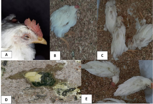
Clinical signs observed in the infected group at 4th and 5th days post- infection (dpi). A, foamy conjunctivitis and periorbital edema were observed at 4th dpi; B, neurological signs (leg paralysis) were observed at 4th dpi; C, sudden high mortalities were observed at 4th dpi; D, greenish lose droppings were observed at 5th dpi; E, neurological signs (torticollis) were observed at 5th dpi.
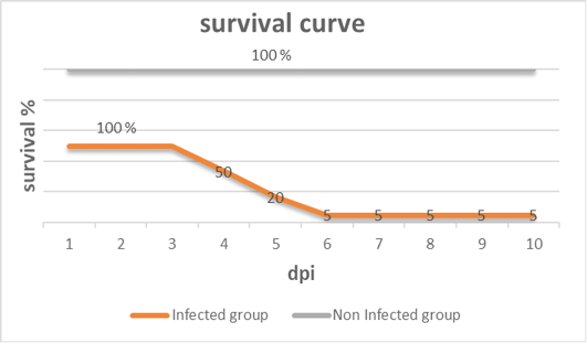
Survival curve after inoculation of 28 days old chickens with vNDV (infected group) and non-infected group of the experiment at different days post- infection (dpi).
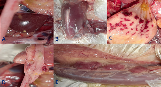
Gross pathological lesions observed in the infected group at 4th and 5th days post- infection (dpi) up on postmortem examination. A, Enlarged mottled spleen; B, severe muscle congestion; C, severe hemorrhages on the tips of periventricular glands; D, hemorrhagic ulcers on cecal tonsils; E, severe congestion with hemorrhages on the thymus gland.
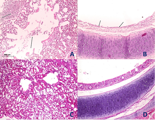
Histological section of lung and trachea of the infected and non-infected group at 5th day post infection (dpi). A, lung showed moderate pneumonia with inflammatory exudate inside tertiary bronchi and secondary bronchi (arrow) with necrosed tertiary bronchi lining; B, trachea revealed tracheitis with noticeable necrosis of lining epithelium and mononuclear cells infiltration (arrow); C, normal lung (non-infected group) with no histopathological alterations; D, normal trachea (non-infected group) with no histopathological alterations.
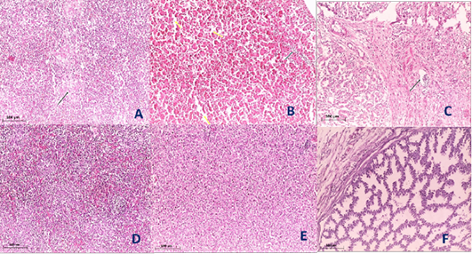
Histological section of spleen, liver and Proventriculus of the infected and non-infected group at 5th day post infection (dpi). A, spleen showed spleenitis with marked focal necrosis of lymphoid tissue (arrow); B, liver showed hepatitis with mononuclear cells infiltration (arrow) and dilatation of hepatic sinusoids (yellow arrow); C, Proventriculus suffered from moderate proventriculitis with mononuclear inflammatory cells infiltration within tunica muscularis (arrow) and epithelial cells degeneration; D, normal spleen (non-infected group) with no histopathological alterations; E, normal liver (non-infected group) with no histopathological alterations; F, normal Proventriculus (non-infected group) with no histopathological alterations.


