Advances in Animal and Veterinary Sciences
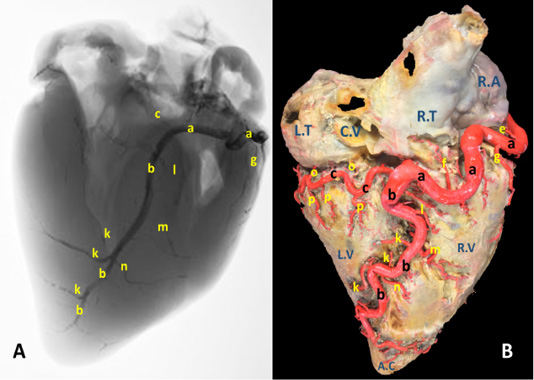
A) A radiograph and B) latex injection of donkey heart showing the distribution of the right coronary artery and its branches on the atrial surface.
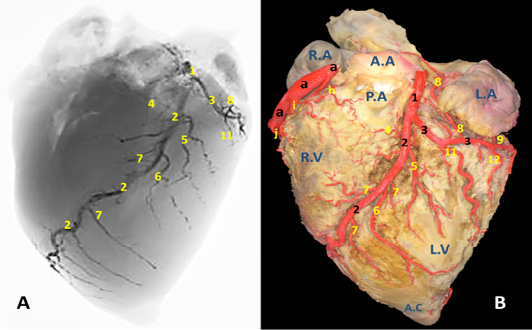
A) A radiograph and B) latex injection of donkey heart showing the distribution of the left coronary artery and its branches on the auricular surface.
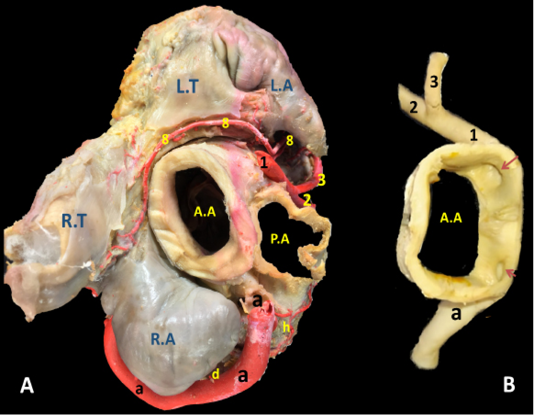
The cardiac base of donkey heart showing A) the origin and directions of the coronary arteries and B) the position of the right and left coronary ostia inside the aorta.
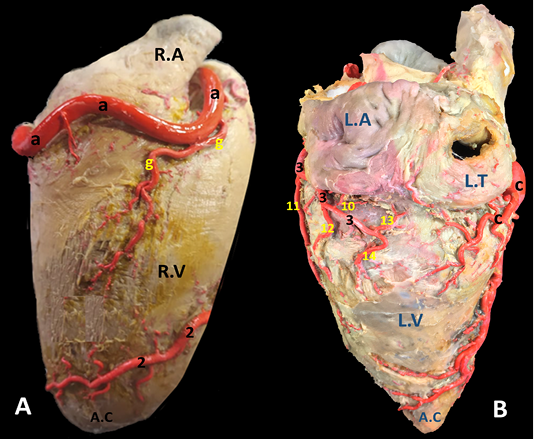
A) Margo ventricularis dexter and B) Margo ventricularis sinister of the donkey heart showing the distribution of coronary arteries.

The auricular surface of donkey heart showing A) ramus diagonalis sinister of a. coronaria sinistra and B) myocardial bridge of r. interventricularis paraconalis.


