Advances in Animal and Veterinary Sciences
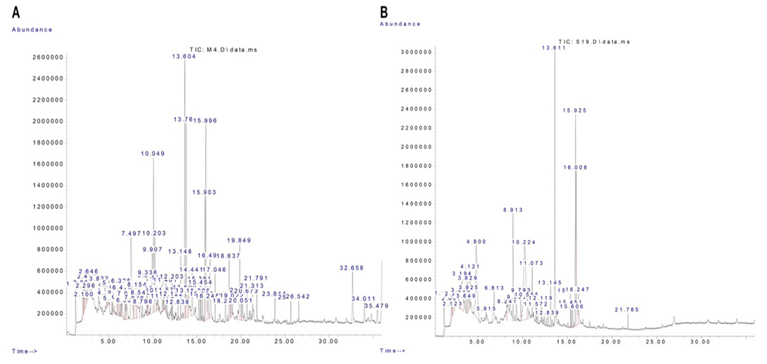
Chromatogram of the methanolic extract of R. epapposum (A) and Lycium shawii (B).
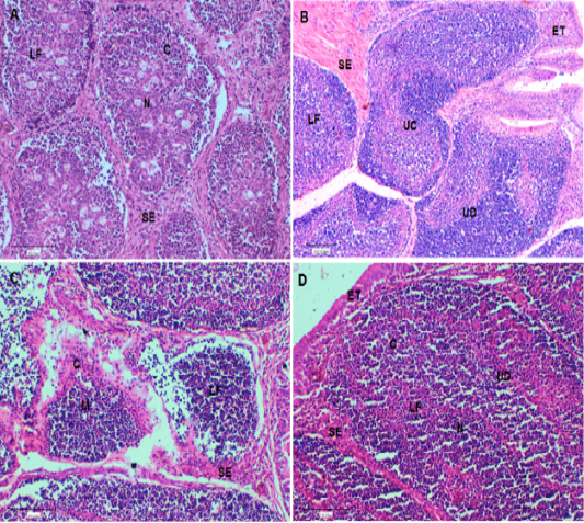
(A). Bursa of control chickens on day 42 showing lymphoid follicles with scattered evacuation and medium condensation of lymphocytes in cortical regions. The demarcation between cortex and medulla was disappeared. (B). Bursa of challenged chickens with sheep erythrocytes (SRBC) on day 42 showing depletion of medulla from lymphocytes with smaller lymphoid follicles. (C). Bursa of chickens challenged with SRBC post-treatment with Lycium shawii showing depletion of cortex from lymphocytes with large empty spaces. (D). Bursa of chickens challenged with SRBC post-treatment with R. epapposum showing condensation of cortex with lymphocytes and medulla with extensive population of lymphocytes with clear demarcation between them. H and E (X40). SI: Space interfollicular, C: Cortex, M: Medulla, LF: Lymphoid Follicle, ET: Epithelial Tissues, UC: Undifferentiated Cells, BC: Blood capillaries.
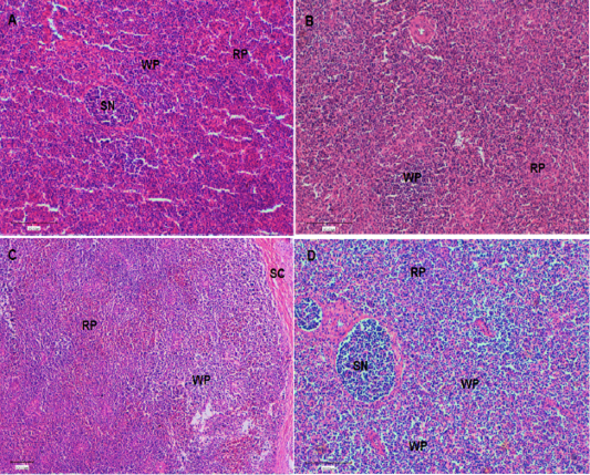
(A). Spleen of control chickens on day 42 showing normal splenic histology and architecture. In addition, different types of red pulp and white pulp were easily visible. (B). Spleen of challenged chickens with sheep erythrocytes (SRBC) on day 42 showing decrease of red pulp with a few white pulp. (C). Spleen of chickens challenged with SRBC post-treatment with Lycium shawii on day 42 showing mild increase in white pulp. (D). Spleen of chickens challenged with SRBC post-treatment with R.epapposum on day 42 showing sever increase in white pulp and condensation of lymphocytes. Differentially types of splenic nodules were easily visible. HandE (X40). RP: red pulp; WP: white pulp, SN: splenic nodule; Sc: splenic capsule.


