Advances in Animal and Veterinary Sciences
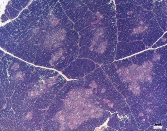
Thymus of broiler chickens in the control group. Stained with hematoxylin and eosin.

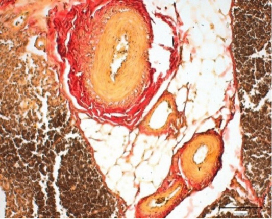
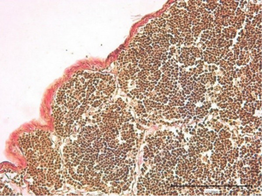
Thymus of broiler chickens in the control group. Blood vessels. Stained by Van Gieson.
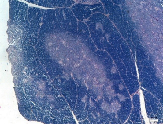
Thymus of the experimantal chickens (Nucleostim, 10 g/kg of feed). Stained with hematoxylin and eosin.
Thymus of the control chickens (Nucleostim, 10 g/kg of feed). The growth of loose fibrous connective tissue. Stained by Van Gieson.
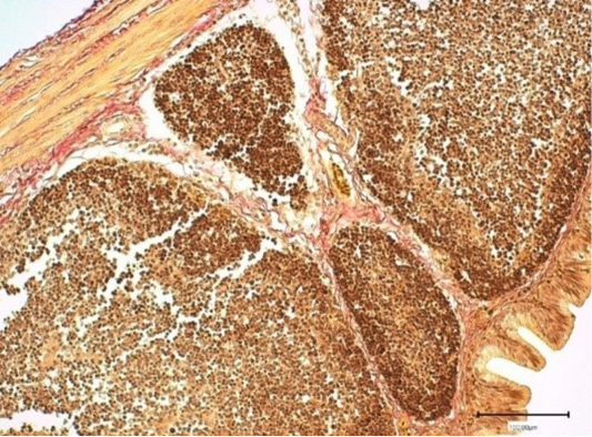
Thymus of the control broiler chickens. Connective tissue capsule. Stained by Van Gieson.
Thymus of the control chickens (Nucleostim, 10 g/kg of feed). Connective tissue capsule. Stained by Van Gieson.

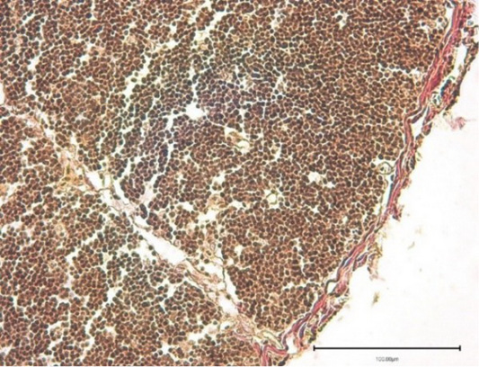
The bursa lobule (lymphoid follicle) of the control broiler chickens. Stained with hematoxylin and eosin.
The bursa fold stroma of cross-breed ROSS308 broiler chickens in the experimental group (Nucleostim, 10 g/kg of feed). Stained by Van Gieson.


