Advances in Animal and Veterinary Sciences
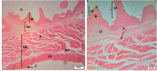
Representative photomicrographs of the rumen in sheep. A(H&E, 10X):M-Mucosa, Sm-Submucosa, ME-Muscularis Externa, IL- inner layer, OL- outer layer, MP- Myenteric plexus, S-Serosa; B(H&E, 40X): E-Epithelium, lp-lamina propria, SP- short papillae, LP- long papillae, Cf- Collagen fibers, V-Vessels.
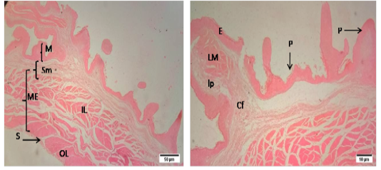
Representative photomicrographs of the reticulum in sheep. A(H&E, 10X): M-Mucosa, Sm-Submucosa, ME-Muscularis Externa, IL- inner layer, OL- outer layer, S-Serosa. B(H&E, 10X): E-Epithelium, lp-lamina propria, P- papillae,, Cf- Collagen fibers, V-Vessels.
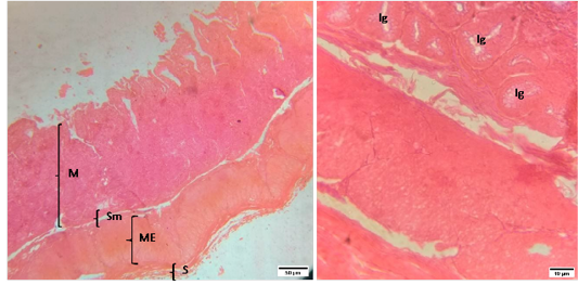
Representative photomicrographs of the cecum in sheep. A(H&E, 10X): M-Mucosa, Sm-Submucosa, ME-Muscularis Externa, S-Serosa. B(H&E, 40X): Ig-Intestinal glands.
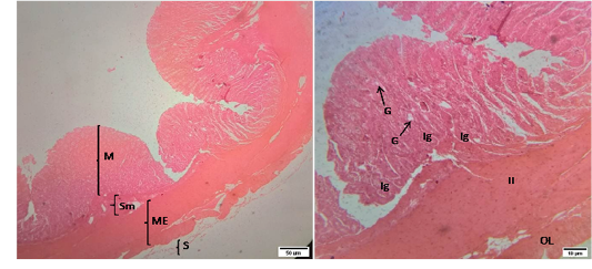
Representative photomicrographs of the colon in sheep. A(H&E, 10X): M-Mucosa, Sm-Submucosa, ME-Muscularis Externa, S-Serosa. B(H&E, 40X): G-Goblet cell, Ig –Intestinal glands, IL-Inner circular muscularis externa, OL- Outer longitudinal muscularis externa.
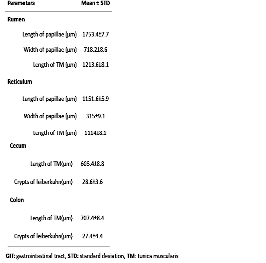
Biometric properties of the GIT i.e. rumen, reticulum, cecum and colon of indigenous sheep (n=5).


