Advances in Animal and Veterinary Sciences

Clinical signs of avian pox in chickens and pigeon. (A, B, C) Nodular lesions of the comb, wattle, around the eyes and ears of the chickens. (D) Nodular lesions on the eyelids and beak of the pigeon. (E) Fibronecrotic lesion on oral mucosal membrane of pigeon.

Pock lesions (red circle) and thickening of CAM of avian pox infection. The pock lesion is thickened, rounded, white colour, 3-5 mm in diameter, and focal.
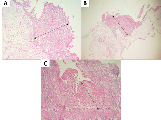
(A, B) Proliferation of chicken stratum spinosum cells (acanthosis) (oblique arrow). (C) Proliferation of pigeon stratum spinosum cells (acanthosis) (oblique arrow) (H and E staining, x100).
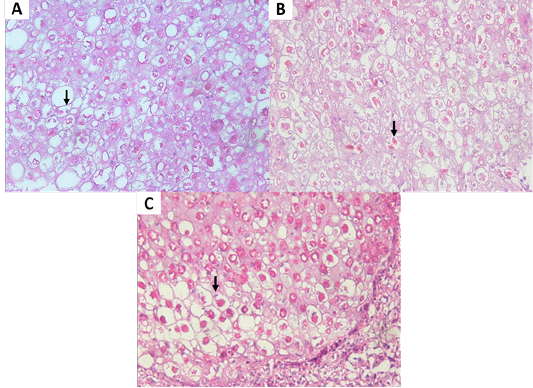
(A, B) Eosinophilic intracytoplasmic inclusion body (vertical arrow) of chicken. (C) Eosinophilic intracytoplasmic inclusion body (vertical arrow) of pigeon. (H and E staining, x400).
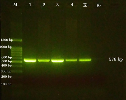
PCR results. (M) 100-bp DNA ladder. (1) chicken a. (2) chicken b. (3) chicken c. (4) pigeon. (K+) positive control. (K-) negative control.


