Advances in Animal and Veterinary Sciences
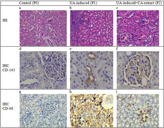
Cross-section of glomerulus and tubulointerstitial of rats showed no change in the structure of the epithelial and mesangium in the control group (a). However, there were severe infiltration cell, vacuolar degeneration, and necrotic epithelium in the area of glomerulus and interstitial tubules on induction group (b) meanwhile decrease the number of inflammation and alteration epithelial cells were observed in the histopathological cross-section of the renal tissue of rats in the group that received Coleus amboinicus. (c). No specific expression of macrophage CD-68 nor positive cells of macrophage CD-163 in the control group (a.d.) Expression of macrophage CD-163 positive cells is observed in interstitial and tubules (e) but decreased numbers of CD-163 expressions in the group with received CA extract (f). Meanwhile, the expression of macrophage CD-68 is also observed an increase in the area epithelial tubularcells in the treatment group (h) and decreased number of positive cells in the group with Coleus amboinicus extract (i) (40 x; IHC, counterstained hematoxylin).


