Advances in Animal and Veterinary Sciences
Research Article
Adv. Anim. Vet. Sci. 9(9): 1483-1488
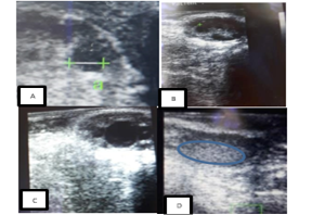
Figure 1
Ultrasound images of a cow’s ovaries in this study. (A) This image shows to get diameter of follicle or corpus luteum, (B) these images show five follicles, (C) these images show one dominant follicle, and (D) these images show of corpus luteum.
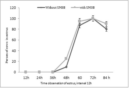
Figure 2
Percentage of estrus response after second injection of PGF2α in cows observed at 12h intervals (with UM3B or without UM3B).


