Advances in Animal and Veterinary Sciences
Research Article

A Comparative Study between the Effect of Synthetic and Natural Propolis on Rabbits skin Wound Infected by Staphylococcus aureus
Zainab J. Malik1, Ali I. Al-ameedi2*, Zahraa M Ayad3, Maymunah Abdullah1
1Department of Surgical and Obstetric, Veterinary Medicine collage, Al- Qasim Green University-Iraq; 2Department of physiology and pharmacology Veterinary Medicine collage, Al- Qasim Green University-Iraq; 3Department of Nursing, Altoosi University College-Najaf-Iraq.
Abstract | Objective: The main objective of this work was to measure the impact of synthetic and natural propolis on infected wounds contaminated with pathogenic staphylococcus. aureus. Material and methods: Sixteen healthy local rabbits were classified into two similar group. All rabbits were anaesthetized by 2% lidocaine hydrochloride S/c as local anesthetic, then surgical incision was performed 3*4cm in the skin. From one day of wound infected with staph. Aureus, first 8 rabbits treated with 5% natural propolis while the animals from (9-16) treated with synthetic propolis, Results: Temperature, respiratory rate and heart rate were included in clinical criteria investigation. there were no significant (p ≤0.05) differences between groups. Macroscopic finding showed that the scar present greater quantity in the animals exposed to synthetic propolis than that in group one furthermore the histopathological findings exhibited that the wound healing in the treated group by natural propolis was quicker and better and the epithelial cells with more maturity so the skin back up near normal, while the synthetic propolis group showed early epithelization with mild regeneration of adnexa. Conclusions: The healing properties of propolis very encouraging to make more trials in different species and types of tissues furthermore to analysis of constituent of propolis by accurate methods.
Keywords | Propolis, Wound, Skin, Rabbit, Staphylococcus. Aureus
Received | April 23, 2021; Accepted | May 06, 2021; Published | July 01, 2021
*Correspondence | Ali I Al-ameedi, Department of physiology and pharmacology Veterinary Medicine collage, Al- Qasim Green University-Iraq; Email: ali.alameedy89@yahoo.com
Citation | Malik ZJ, Al-ameedi AI, Ayad ZM, Abdullah M (2021). A comparative study between the effect of synthetic and natural propolis on rabbits skin wound infected by staphylococcus aureus. Adv. Anim. Vet.Sci. 9(8): 1233-1237.
DOI | http://dx.doi.org/10.17582/journal.aavs/2021/9.8.1233.1237
ISSN (Online) | 2307-8316; ISSN (Print) | 2309-3331
Copyright © 2021 Al-ameedi et al. This is an open access article distributed under the Creative Commons Attribution License, which permits unrestricted use, distribution, and reproduction in any medium, provided the original work is properly cited.
Introduction
The scientific community still busy lately in searching for natural plant and animal preparations for the purpose of using them in the treatment of bacterial, fungal and viral infections to get rid of the problems of adverse effects of drugs and reduce the resistance of organisms. Propolis is the common name of a combination of resinous compounds obtained in the northern temperature region by honeybees from sections of trees, buds and exudates, ranging from the chestnut are the primary origins of propolis (Wael et al., 2015). In the hive, propolis is typically used to cover the inner walls, to shield intruders, such as lizards and snakes, or even against wind and rain, and to discourage the growing of fungi and bacteria (Zhang, et al., 2009). It also forms phytochemical components proven to support differentiation or apoptosis in cells (Martinotti S and Ranzato E, 2014) Caffeic acid, caffeic phenyl ester, artepillin C, Quercetin, resveratrol, galangin, and genistein are among other Propolis, along with other honeybee commodities has potent therapeutic effects and has been used in overall prevalence in different countries since ancient times (Kuropatnicki et al., 2013). It is thought that propolis has antiseptic, antibacterial, antimycotic, astringent, spasmolytic, anti-inflammatory, anesthetic, antioxidant, antifungal, anti-ulcer, anticancer and immunomodulatory impact (Sawicki et al., 2012; Martinotti et al., 2015) Honey is a nutritious, well balanced food and has been considered an important cure for many diseases (Jull et al., 2015). Unfortunately, the phytochemical properties of the varieties of honey used in wound healing experiments of both human and veterinary medicine are not recorded.
Materials and methods
Hydrochloride lidocaine: 2% B.P. Netherlands, Holden, Lelystad
• Streptomycin - Penicillin (Penoksa LA, Vilsan Ankara).
• Using the silicone propolis (capsule): open the capsule and place the gelatine in it.

Figure 1: Propolis capsule
Propolis Ethanolic Extraction
The technique according to (Al-Ameedi and Nahi 2019; Paviani et al., 2012). Used in which 3 g of crude propolis was blended with 10 ML of ethanol (Merck, Darmstdadt, Germany) and stirred using a magnet stirrer for one day at room temperature using the ethanolic extract from green and Red propolis. The insoluble component was then filtered out. The filtrates were kept in a freezer overnight at 10 ° C, then filtered again to decrease the extracts’ wax content. Solvent evaporated to dry ethanolic extract of green and red propolis at the temperature of 60 ° C in a vacuum oven, and the production findings have been focused on the original propolis quantity.

Animals
Sixteen adult rabbits from the College of Veterinary Medicine / AlQasim Green University’s Herd Animal Resource were chosen. The Rabbit were aged (3 10) months and weighted (1,5-2) Kg, housed for surveillance and adaptation in controlled surroundings, followed by two (50*70) cm storage cages for all of the experiments.
Experimental design
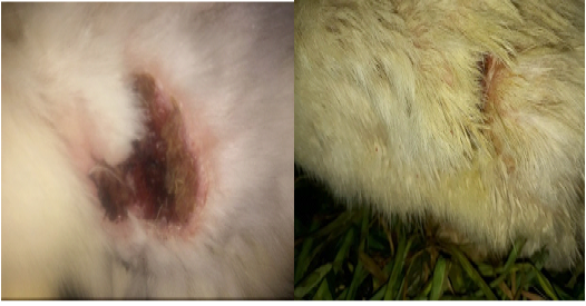
At once and after induced skin wound, contaminated this wound with 1.5 x 10⁷ CFU / ml of staph. aureus in physiological saline were obtained from microbiology lab of college of veterinary medicine at Al-Qasim green university.
Ointment preparation: according to (Al-Ameedi and Nahi 2019).
First group: group (A): In this group animals were numbered (1- 8), have skin wound 3*4 cm was treated with artificial propolis (one capsule about 3g).
Second group (B): Throughout this group were numbered (9-16), skin wound 3*4 cm were covered with natural propolis that processed into cream (3g). During the 1st Week and 3rd Week, histopathology was taken.
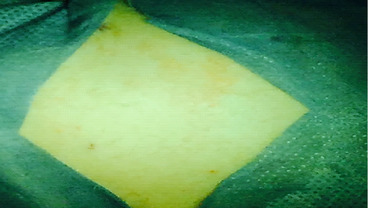
Figure 3: Preparation the site of operation
Clinical observation
Temperature, respiratory rate and heart rate, defecation and urination were medically and clinically tested for one week after surgery.
Histopathological tests
On 15th and 30th day after surgery, skin biopsies were performed. Biopsy is fixed in 10% formalin neutral buffer, then treated regularly and integrated with paraffin as a block cut at a height of five to six micrometers and stained with Hematoxylin and Eosin dye (Luna,1968).
Study Statistical
The outcome was seen as mean + S.E. Parametric results were evaluated using two approaches, which were contin
Table 1: Effect of synthetic and natural propolis on body temperature (Fo), respiratory rate and heart rate (Mean ± SE).
| Days | Temperature (Fo>) | Respiratory rate | Heart rate | |||
|
Nature P. Mean±S.E |
Synthetic P. Mean±S.E |
Nature P. Mean±S.E |
Synthetic P. Mean±S.E |
Nature P. Mean±S.E |
Synthetic P. Mean±S.E |
|
|
1 |
103.54±3.07 b |
105.20±4.97a | 71.50±3.30a | 69.71±3.82a | 165.6±5.34a | 169.50±2.56a |
|
2 |
105.32±3.81b | 104.33±5.11a | 55.62±3.51b | 70.45±3.71a | 147.20±6.53a | 157.10±2.43b |
|
3 |
105.10±3.72b |
106.00±5.71a |
50.37±4.12b |
54.25±2.92b | 143.25±2.94a | 145.60±3.19b |
|
4 |
98.31±6.70a | 101.32±3.76b | 41.50±2.28b | 43.37±4.32b | 139.10±1.51b |
131.80±3.50b |
|
5 |
100.21±4.45a |
108.31±9.10b |
43.62±2.89b | 43.21±2.87a | 136.62±1.54b | 133.45±2.57b |
|
6 |
104.51±3.20a |
108.10±8.34a |
40.25±2.37a |
43.75±2.50a | 140.01±1.94b | 138.20±2.13b |
|
7 |
103.33±4.90a | 99.84±6.91a | 42.87±2.56b | 46.37±3.31b | 133.10±2.01b |
140.62±1.73a |
Different small letter denotes there is no significant (p≤0.05) between group.
ued with LSD and were considered to be important, of variance analysis (ANOVA). The Social Sciences Statistics Kit (SPSS, 2010) has been used (SAS, 2001).
Results and discussion
Signs outcome
Temperature, respiratory rate, cardiac rate, defecation and urination of the First week after intervention found increased, with regular defecation and urination in all the animals, only three animals in natural propolis and one animal in the synthetic propolis group. Significant overlap of findings in the after-day control group and the treated group but early disappears in the natural propolis and skin treatment groups as opposed to the third day, synthetic propolis treatment group 5 to 7 days after operation, which may be attributed to a rise in the blood flow in the surgical site. Apart from the rise in blood vessel dilatation and improved capillary permeability, other researchers accepted (Lu et al., 2017). that these clinical elements had no major improvements before and after operation.
Discussion
Natural antioxidant plays a vital role in improving the immune system and restoring the normal functions of the body, propolis one of these antioxidants generally produced from plant by worker bees. The present study exhibited that propolis increase the time of closer of incision due to restore and increase the production of collagen type 1 at the site of incision. Furthermore, a direct inhibitory effect of propolis on cytokine production by immune cells has been documented (Agren et al., 2015). The wound repair-enhancing effects of propolis are partly due to its anti-inflammatory properties. It is thought that prolonged inflammation impairs the healing process (Bufalo et al., 2014). The sum of scar suggested the efficiency of wound healing, and the macroscopic examination findings revealed more scar tissue forming in the synthetic propolis than in other groups at the seventh day after surgery. While the scar tissue in the synthetic propolis community fallen during the seventh day after surgery, the minimal amount of scar tissue that may be triggered by pathways in the 4 forms of healing decreased (Gallo et al., 2014). Primary cure happens when a wound is healed within hours of being developed. Main delayed healing takes place when a wound is left open intentionally for a certain duration before closing. Secondary purpose care happens with or without topical medication with wounds that are left to curate (Tan et al., 2012) Here, dressing Adjustments are carried out before contraction and epithelialization shut down the wound. Finally, epithelialization of the partial thickness wounds or the epidermis and part of the dermis (Glat et al., 1997). Implementation trauma is one of the most Important stimuli to initiate the development of an inflammatory response. Routine surgical procedures require various stages of tissue management, which can cause inflammatory responses which eventually lead to adhesion Forming by initial tissues abrasion, desiccation, ischemia, bleeding, infection and exposure to foreign materials (Liakakos et al., 2001). Fibroblasts are associated with adhesion throughout the day from five to ten, while collagen deposition and organization progress. Fibroblasts are predominantly the comparatively few cells present. The collagen fibrils are arranged into distinct bundles interposed by fibrocytes and a few macrophages 7 to 21 days after injury. Sometimes protected by mesothelium, large well-defined adhesions include blood vessels and connective tissue fibers (Dizerega, 1994; Alizzi, 2005)

Pathological study
1-Group one by synthetic propolis:
At 7th Day Post operation:
The histopathological examination in treated group at 7th day Post operation showed there was early epithelization with moderate restoration of adnexa with scattered eosinophilichylinized fiber (Fig.8).
At 21th Day post operation:
The histopathological examination in treated group at 21th day Post operation showed moderate epithelization good restoration of adnexa with persistence deposition of thick eosinophilic collagen fibers (Fig.9).
2-Group tow by natural propolis:
At 7th Day post operation:
Histopathological sections of treated group at 7th day post operation, showed good epithelization with small amount of edema in the surface the granulation tissue less cellular well vascularized (Fig.10). Another section showing high magnification of granulation tissue showing proliferation of fibroblast and myofibroblast (Fig.11) At 21th Day post operation: Histopathological sections of treated group at 21th day post operation, showed excellent epithelization and remodeling like to normal tissue with keratin formation apoptosis in keratinocyte also seen (Fig.12). Good epithelization with early granulation tissue formation characterized by highly cellular neovasculasation and myofibroblast also seen. The natural propolis group obtained the highest group in histopathological findings and the best wound healing; this finding is believed to contribute to the healing properties of propolis because of the different mechanisms, including propolis› antimicrobial properties, primarily due to the flavonoid content and in particular the presence of pinocembrin, galangan, and pinobanksin. Antifungal effects are also demonstrated in Pinocembrin. Ester coumaric and caffeic Acids are other chemicals with well-established effects (Wael et al., 2015; Iftikhar et al., 2010).
Conclusion
In conclusion the healing properties of propolis very Encouraging to make more trials in different species and types of tissues furthermore to analysis of constituent of propolis by accurate methods.
Acknowledgments
The authors grateful to all the Staph of Departments of physiology and pharmacology, College of Veterinary Medicine at Al-Qasim Green University for their help and support.
Conflict of interest
There is no conflict of interest.
Authors Contribution
All authors are involved equally in the work and writing of this article and approved it in publication
References


