Advances in Animal and Veterinary Sciences

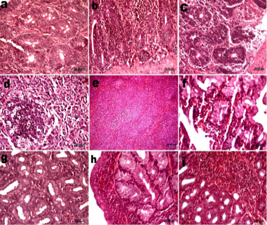
Histological sections of trachea, kidneys and proventriculus of vaccinated groups (a) Trachea group 2 at 5dpv showing inflammatory cells infiltration in lamina propria. (b) Trachea group 3 at 2dpv showing mucous in surface epithelium. (c) Trachea group 3 at 15dpv showing lymphocyte infiltration. (d) Kidneys group 2 at 2dpv showing mild edema in interstitial tissue and hemorrhage. (e) Kidneys group 3 at 2dpv showing foci of degeneration in renal tubules. (f) Kidneys group 3 at 5dpv showing degeneration in renal tubules and inflammatory cells infiltrations. (g) Kidneys group 3 at 10dpv showing heavy infiltration with lymphocytes. (h) Proventriculus group 2 at 5dpv showing mononuclear inflammatory cells infiltrations. (i) Proventriculus group 3 at 5dpv showing increased gland secretions which accumulated in the lumen.
Histological sections of small intestine, spleen and Harderian gland of vaccinated groups (a) Small intestine groups 2 at 2dpv showing hyperplasia in intestinal glands. (b) Small intestine groups 3 at 10dpv showing infiltration with lymphocytes. (c) Small intestine groups 2 at 15dpv showing lymphocyte infiltrations. (d) Spleen groups 2 at 2dpv showing proliferation in lymphoid follicles. (e) Spleen groups 2 at 10dpv showing Activation of lymphoid follicles. (f) Harderian gland groups 2 at 2dpv showing hemorrhage. (g) Harderian gland groups 3 at 5dpv showing lymphocytes infiltrations. (h) Harderian gland groups 2 at 10dpv showing lymphoid follicles. (i) Harderian gland groups 3 at 15dpv showing lymphoid follicles.


