Advances in Animal and Veterinary Sciences
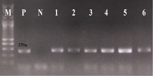
The results of conventional PCR technique, using the primer pairs ParvoInt2FB/ ParvoInt2CR: the positive reaction indicated by an amplicon that is 275 bp in length (M: DNA ladder 100bp, P: positive control (Primodog, Merial, France), N: negative control (PCR grade water), 1-6: positive samples obtained in this study).
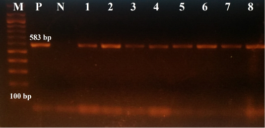
The results of conventional PCR technique, using the primer pairs 555for/555rev: the positive reaction indicated by an amplicon that is 583 bp in size (M: DNA ladder 100bp, P: positive control (Primodog, Merial, France), N: negative control (PCR grade water), 1-8: positive samples obtained in this study).
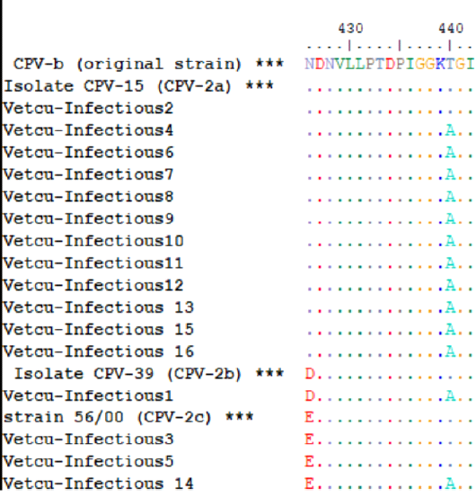
The multiple amino acid alignments of the Egyptian sequences with reference strains for CPV-2 antigenic typing; the Egyptian sequences (Vet Cu-Infectious 1-16, accession numbers: MT596684-MT596696 and MT636872-MT636874) were aligned together with reference sequences (asterisks) that include [CPV-b (original strain, M38245.1), CPV-15 (CPV-2a, M24003.1), CPV-39 (CPV-2b, M74849.1) and strain 56/00 (CPV-2c, FJ222821.1)], which indicates that CPV-2a was the predominant variant.


