Advances in Animal and Veterinary Sciences
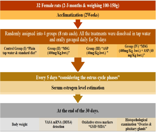
Experimental plan
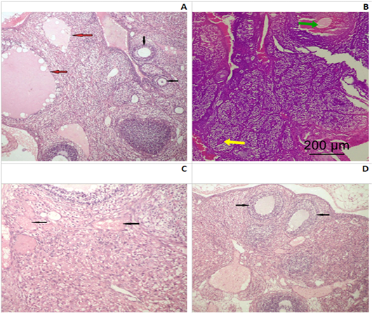
Photomicrograph of an ovarian section of adult Albino rats: (A) Control group I, the ovaries possessed normal histological architecture structure; well formed mature secondary follicles (red arrows) alternating with small primary follicles (black arrows). (B) Group II (SMG) showed less mature graffian follicles (yellow arrow) and congested blood vessels (green arrow). (C) Group IV (SMG +ASP) showed numerous markedly congested and dilated blood vessels with diffused hemorrhagic areas (black arrows). (D) Group IV (SMG +ASP) showed lower number of ovarian follicles with less maturity of the secondary follicles (black arrows) (H&E x200).
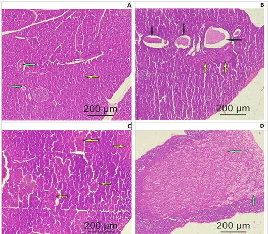
Photomicrograph of a pituitary gland section of adult female Albino rats: (A) Control group I showing normal histological structure of the secretory cells (yellow arrow) and blood vessels (green arrow) (B) Group II (SMG) show cystic formation (black arrows) and few fibrotic bands (yellow bands). (C) Group III (ASP) showing degenerated and necrotic areas with fragmented nuclei (yellow arrows). (D) Group IV (SMG +ASP) showing large fibrotic area (green arrow) involving most of the pituitary anterior lobe (yellow arrows) (H&E x200).


