Advances in Animal and Veterinary Sciences
Research Article
Adv. Anim. Vet. Sci. 9(5): 692-699

Figure 1
Photograph showing the gastrointestinal tract of crocodile.
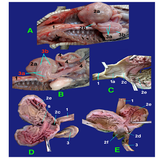
Figure 2
Photograph showing structures of esophagus and stomach.
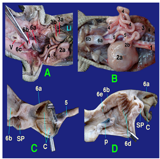
Figure 3
Photograph showing the structures of colorectum
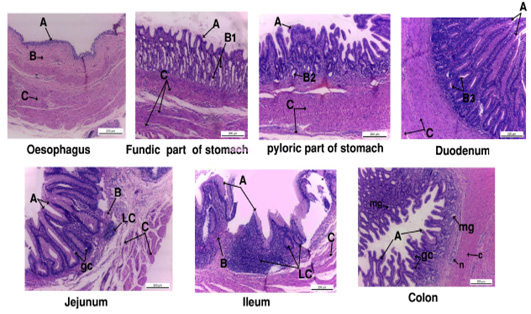
Figure 4
Photograph showing the histology of gastro-interstinal tract.
A, tunica mucosa ; B, tunica submucosa; B1, fundic gland; B2, pyloric gland; B3, duodenal gland; gc, goblet cell ; LC, lymphocyte; mg, mucous gland.
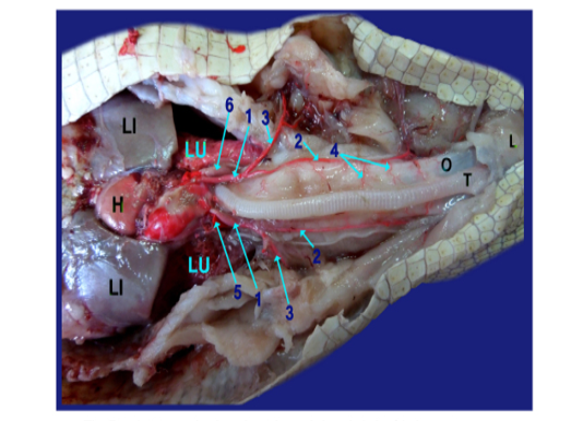
Figure 5
Photograph showing the origin of the right and left aorta.
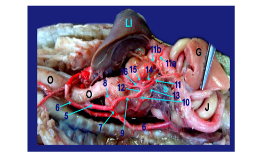
Figure 6
Photograph showing the branches of celiacomesentric artery.
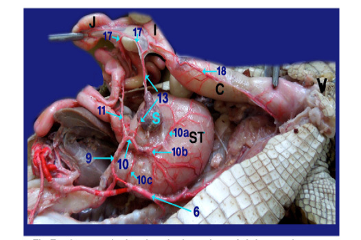
Figure 7
Photograph showing the branches of right gastric artery.
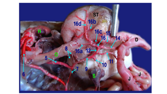
Figure 8
Photograph showing the branches of gastroduodenal artery.
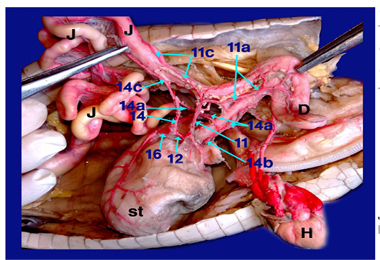
Figure 9
Photograph showing the arterial supply of duodenum and jejunum.


