Advances in Animal and Veterinary Sciences
Case Report
Adv. Anim. Vet. Sci. 9(5): 689-691
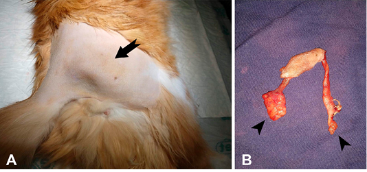
Figure 1
Trichogranuloma in a cat. A. Right pelvic limb showing a firm elastic nodular mass (arrow). B. Surgical excision of the nodular mass showing two tubelike structures (fistulous tract) with blind edges (arrow heads).

Figure 2
Trichogranuloma in a cat. Cutaneous nodule. There are fragments of free hair shaft (arrows) surrounded by giant foreign body cells (arrow heads) and leukocytic infiltration. (400 X, H&E).


