Advances in Animal and Veterinary Sciences
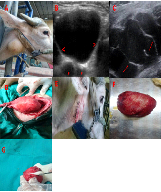
A) Branchial cyst in a 2 years-old buffalo just behind the mandibular angle. B) and C) ultrasonography of brachial cyst wall (>), bifurcation of common carotid artery (*) and septa (arrow). D) blunt dissection of the cyst from the surrounding structure. E) suture of the skin after the end of surgery. F) the cyst after removal. G) milky fluid from the cyst after excision.
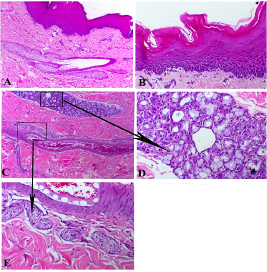
Branchial cyst, A) showed stratified epithelial lining with sub-epithelial sebaceous glands, X100. B) papillary-like stratified squamous epithelium, X200. C) sub-epithelial glandular, vascular and neuronal appendages (arrows indicates insets of both mucous glands and nerve bundles), X 100. D) mucous glands, X200. E) neuronal structure around the blood vessels, X200. H&E stain.

A) cystic structure filled with serous exudates (arrowheads indicates exudate and arrow indicates fibrous capsule of the cyst rich with fibrin bands, X100. B) haemorrhagic cyst showed intensive haemorrhage with organization attempts at the border of the cysts papillary-like stratified squamous epithelium, X100. H&E stain.


