Advances in Animal and Veterinary Sciences
Research Article

Anatomical Studies on the Arterial Blood Supply of the Pelvic Limb of Geese
Meray Nabil Ramsis1, Samar M. EL-Gammal1*, Khaled Abo-EL-Sooud2, Gamal A. Swielim1
1Anatomy and Embryology Department, Faculty of Veterinary Medicine, Cairo University, Giza, Egypt; 2Pharmacology Department, Faculty of Veterinary Medicine, Cairo University, Giza, Egypt.
Abstract | The present study was conducted on 25 adults, healthy Egyptian native breed of geese. The arteries of the pelvic limb were demonstrated by injection of colored gum milk latex and treated by the ordinary method of preservation. The arterial vascularization of the pelvic limb was mainly obtained from the external iliac and ischiatic arteries. The external iliac artery supplies the pelvic limb to the level of the knee joint, while the ischiatic artery is responsible for supplying the entire limb, changing its name according to the region of the limb it supplies. The ischiatic artery terminates after giving off the sural artery and continues in the leg region as the popliteal artery. The branches of the popliteal artery supply the knee and leg regions; whereas the cranial tibial artery, the continuation of the popliteal artery, supplies the foot with its own branches. The presence of extensive arterio-venous anastomosis (rete tibio tarsale) was shown to clarify means of thermoregulation in limbs. The aim of the present work is to highlight the accurate angio-architecture of the pelvic limb which is pivotal for surgical interference in cases of joints, limb and foot affections in water birds.
Keywords | Pelvic limb, Geese, External iliac, Ischiatic, Arterial supply.
Received | Deccember 08, 2020; Accepted | December 17, 2020; Published | February 20, 2021
*Correspondence | Samar M. El-Gammal, Anatomy and Embryology Department, Faculty of Veterinary Medicine, Cairo University, Giza, Egypt; Email: samarelgammal13@yahoo.com
Citation | Ramsis MN, El-Gammal SM, Abo-El-Sooud K, Swielim GA (2021). Anatomical studies on the arterial blood supply of the pelvic limb of geese. Adv. Anim. Vet. Sci. 9(4): 604-614.
DOI | http://dx.doi.org/10.17582/journal.aavs/2021/9.4.604.614
ISSN (Online) | 2307-8316; ISSN (Print) | 2309-3331
Copyright © 2021 El-Gammal et al. This is an open access article distributed under the Creative Commons Attribution License, which permits unrestricted use, distribution, and reproduction in any medium, provided the original work is properly cited.
INTRODUCTION
Poultry meat production is a worldwide business that progressively has become the standard form of mass production of protein (Jordan and Pattison, 1996). Despite this, geese have never been exploited commercially as much as chickens or even ducks have been (Chelmonska, 1995). Domestic geese were widely farmed to provide a source of meat, livers, eggs, and feathers. Historical records point to the use of geese, e.g., the fattening of goose for the table, and force-feeding has been known since ancient Egyptian times (Johanna et al., 2018). Bird’s foot was exposed to injuries, wounds, bumble foot, fibrosis, and swelling of the metatarsal pad, where the most common and needed surgical interference is done (Heidenreich, 1997, Routh, 2000; Cooper, 2002).The importance of the poultry among our native livestock has initiated an increasing interest to establish accurate and detailed anatomical facts about the arterial vascularization of the pelvic limb of the geese (Swielim et al., 2012).
MATERIAL AND METHODS
The present work was carried out on 25 adult healthy Egyptian native breed of geese of both sex. Their average body weight was (2.8-3.5) kg with ages about 120-140 days. They were obtained from local markets and transported to the laboratory of Anatomy and Embryology Department, Faculty of Veterinary Medicine, Cairo University.
The arterial-architectural study
Before exsanguination, the birds were anaesthetized by intramuscular injection of 0.5 ml of xylazine 2% (xylazine hydrochloride, Dutch Farm veterinary pharmaceuticals) then injected by heparin (Cal heparin, 5000I.U) in wing vein for 30 minutes. Each specimen was slaughtered according to the Islamic method of slaughtering through the common carotid arteries and the jugular veins and then left to bleed for 15 minutes, the arteries of injection were cannulated and thoroughly washed by warm normal saline solution Nacl 0.9% containing amount of heparin (heparin calcium 5000 I.U.) to prevent blood clotting and blood remaining in the vessels.
Latex neoprene injection technique
Immediately following euthanasia, the ribs and the sternum were carefully removed to expose the heart. The ventricular apex of the heart was removed and a Nelaton catheter of size 6F to 8F (MA MEDICAL company) was introduced into the left ventricle of the heart to the aorta for injection of the pelvic limb. The injected material was 60% latex neoprene colored with red Rottring ink (Tompsett and Wakelly, 1965). The specimens were then preserved in a container of 10% formalin solution, 2% phenol, and 1% glycerin for 3-5 days at room temperature (25oC) to allow solidification of the latex. The specimens were dissected for exposing the arterial distribution patterns.
RESULTS
The chief arterial supply of the pelvic limb comprises the external iliac and ischiatic arteries.
A. iliaca externa
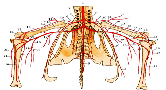
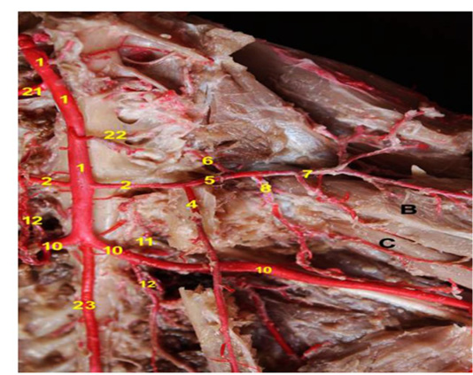
The external iliac artery (Figs.1,2/3&3, 4, 5, 6/2);a short trunk, arises from the lateral side of the descending aorta, about 1cm cranial to the hip joint. It proceeds caudal to the cranial renal artery and passes cranio-laterally within the division between cranial and middle lobes of the kidney. The external iliac arterygivesoff the pubic artery then continues outside the pelvis as the femoral artery through the pre-acetabular notch.
A. pubica
The pubic artery (Figs.1/7& 2/8 &3/3 &4, 5, 6, 9/4) is long slender branch, detached from the lateral aspect of the external iliac artery, 1cm caudal to the origin of the latter artery. It is considered the longest branch of the external iliac artery and proceeds caudo-ventrally with the same curvature of the pubic bone, incinerates itself between the vicinity of the internal obturator and abdominal muscles. During its course, it gives off fine branches that penetrate the cranial part of incisura pubo-ischiadica to supply theM. obturatorius lateralis. The pubic artery continues its course on the ventral border of the pubis to be distributed in the Mm. obturatorius medialis andpubo-ischio-femoralis.
|
1. Aorta descendens |
14. A.renalis media |
|
2. A.mesentrica cranialis |
15. A.renalis caudalis |
|
3. A.iliaca externa |
16. A.caudalis coxae |
|
4. A.ischiadica |
17. A.femoralis caudalis |
|
5. A.iliaca interna |
18.A.nutricia proximalis femoralis |
|
6. A.mediana caudae |
19. A.suralis |
|
7. A.pubica |
20. A.suralis lateralis |
|
8. A.umbilicalis |
21. A.suralis medialis |
|
9. A.cranialis coxae |
22. A.poplitea |
|
10. A.femoralis |
23. A.genicularis proximalis |
|
11. A.femoralis cranialis |
24. A.tibialis medialis |
|
12. A.femoralis medialis |
25. A.tibialis caudalis |
|
13. A.genicularis medialis |
26. A.tibialis cranialis |
A. femoralis
The femoral artery (Figs. 1,2/10 & 3/4 &4, 5, 6/5); represents the direct continuation of the external iliac artery, descends ventro-medially to reach the level of the head of the femur where it detaches dorsally small vessel; the cranial coxal artery, then it bifurcates into the cranial and medial femoral arteries.
A. Cranialis Coxae: The cranial coxal artery (Figs.1/9 &2/7 &4, 5, 6/6); arises from the dorsal aspect of the femoral artery. It releases several branches to supply Mm. ambiens, ilio-tibialis cranialis, ilio-trochantricus cranialis, ilio-femoralis externus, ilio-tibialis lateralis, femoro tibialis externus as well as the capsule of the hip joint.
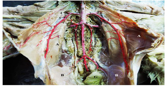
Figure 3: A photograph showing the termination of the descending aorta and the main arteries of the pelvis, (Ventral view).
|
A.M.obliquus externus abdominis |
2. A.iliaca externa |
|
B.M.obliquus internus abdominis |
3. A.pubica |
|
C.Divisio renalis cranialis |
4. A.femoralis |
|
D. Divisio renalis media |
5. A.ischiadicus |
|
E. Divisio renalis caudalis |
6. A.renalis media |
|
F. Ureter |
7. A.renalis caudalis |
|
G. Cloaca |
8. A.iliaca interna |
|
1. Aorta descendens |
9. A.mediana caudae |
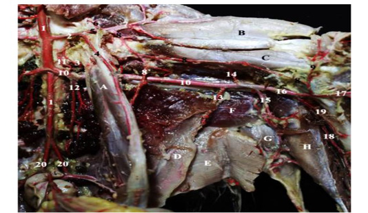
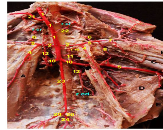
Figure 4: A photograph showing the arteries of pelvic and thigh regions; (ventro-medial view); left side.
|
A. M.obturatorius medialis |
8. A.femoralis medialis |
|
B. M.iliotibialis cranialis |
8`. R.muscularis |
|
C. M.femorotibialis medialis |
9. A.genicularis medialis |
|
D. M.ischiofemoralis medialis |
10. A.ischiadica |
|
E. M.flexor cruralis medialis |
11. A.renalis media |
|
F.M.pubo-ischio-femoralis caudalis |
12. A.renalis caudalis |
|
G. M.gastrocnemius pars lateralis |
13. A.femoralis caudalis |
|
H. M.flexor perforatus digiti III |
14.A.nutricia proximalis femoralis |
|
1. Aorta descendens |
15. A.suralis |
|
2. A.iliaca externa |
16. A.poplitea |
|
3. A.caudalis coxae |
17. A.tibialis medialis |
|
4. A.pubica |
18. A.tibialis caudalis |
|
5. A.femoralis |
19. A.tibialis cranialis |
|
6. A.cranialis coxae |
20. A.iliaca interna |
|
7. A.femoralis cranialis |
|
A. Femoralis cranialis
The cranial femoral artery (Figs. 1,2/11&4, 5,6/7&7, 9/7); emanates from the parent trunk at the level of the hip joint and passes cranio-laterally in the thigh region between the Mm. ilio-tibialis cranialis and femoro-tibialis accessories. It gives off fine collateral branches to supply the preceding muscles as well as the Mm.pectinus and ambiens.
A. femoralis medialis
The medial femoral artery (Figs.1,2/12 &4, 5,6,7/8); long distal branch of the femoral artery, directed caudo-ventrally to the head of the femur where it passes between the neck of the femur and tendon of the M. ilio-femoralis internus in the medial aspect of the thigh. The medial femoral artery proceeds between theMm.ischiofemoralis and femoro-tibialis medialis at the caudal surface of the femur till reach the stifle joint. During its course, it gives off a muscular branch to supply Mm. ilio-femoralis internus, ilio-trochantricus cranialis, ilio-trochantricuscaudalis, femo-tibialis medialis and adductor muscle. It joins the medial tibial artery on the medial aspect of the stifle joint.
|
B. M.iliotibialis cranialis |
8. A.femoralis medialis |
|
C. M.femorotibialis medialis |
10. A.ischiadica |
|
1. Aorta descendens |
11. A.renalis media |
|
2. A.iliaca externa |
12. A.renalis caudalis |
|
4. A.pubica |
21. A.mesentrica cranialis |
|
5. A.femoralis |
22. A.renalis cranialis |
|
6. A.cranialis coxae |
23. Pars synsacralis aortae |
|
7. A.femoralis cranialis |
|
A. ischiadica
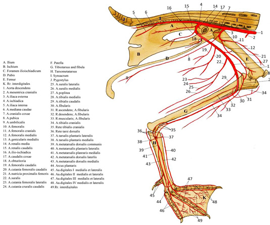
The ischiatic artery (Figs.1, 2/4 & 3/5 & 4, 5, 6, 7, 9/10); the main source of the arterial blood supply of the pelvic limb. It originates from lateral aspect of the abdominal aorta, caudal to the origin of the external iliac artery by about 1.5 cm at the level of 4th - 5th sacral segment of synsacrum. The ischiatic artery dips in the renal fossawhere it gives off its intra-pelvic branches; the middle renal (Figs. 1, 2/14 & 3/6 & 4, 5, 6/11), caudal renal (Figs.1, 2/15, 3/7 & 4, 5, 6/12), obturator (Fig.2/18) and ilioischiatic (Fig.2/16) arteries, then leaves the pelvis via the cranial end of ilioisciatic foramen. The extra-pelvic part of the ischiatic artery proceeds ventro-laterally to the thigh region and becomes incinerated between the vicinity of the M.ilio-fibularis laterally and M. ischio-femoralis medially. It continues distally between M.ilio-tibialis lateralis and M.caudo-iliofemoralis till reaches the caudal aspect of the stifle joint. Along its extra-pelvic course, the ischiatic artery detaches; the caudal coxal, caudal femoral, proximal nutrient femoral, caudal
|
A. M.obturatorius medialis |
10. A.ischiadica |
|
B. M.iliotibialis cranialis |
11. A.renalis media |
|
C. M.femorotibialis medialis |
12. A.renalis caudalis |
|
D. M.ischiofemoralis medialis |
20. A.iliaca interna |
|
E. M.flexor cruralis medialis |
21. A. mesentrica cranialis |
|
1. Aorta descendens |
22. A. renalis cranialis |
|
2. A.iliaca externa |
23. Pars synsacralis aortae. |
|
4. A.pubica |
24. A. mediana caudae |
|
5. A.femoralis |
rcr. Divisio renalis cranialis |
|
6. A.cranialis coxae |
rm. Divisio renalis media |
|
7. A.femoralis cranialis |
rcd. Divisio renalis caudalis |
|
8. A.femoralis medialis |
|
cutaneous femoral and the sural arteries. It continues its course as the popliteal artery in the leg region.
A. caudalis Coxae
The caudal coxal artery (Fig. 1/16, 2/17& 4/3); the first extra-pelvic branch of ischiatic artery, originates from the caudal aspect of the parent trunk at the level of the 4th sacral segment of synsacrum. It bifurcates into medial and lateral branches; the former passes caudo-ventrally to the hip joint, parallel to the shaft of femur where it ramifies in Mm.obturator medialis, ischiofemoralis, pubo-ischio femoralis, flexor cruris medialis, flexor cruris lateralis and caudo-iliofemoralis. The lateral branch supplies the ischiofemoralis and ilio-fibularis muscles then penetrates the latter muscle to supply the M. ilio-tibialis lateralis.
A. femoralis caudalis
The caudal femoral artery (Figs.1/17 &2/19&4/13&7/13); detached from the caudal aspect of the ischiatic artery. It courses caudo-ventrally to the thigh region to supply M.ilio-fibularis and the flexor muscles of the thigh.
|
B. M.iliotibialis cranialis |
10. A.ischiadica |
|
C. M.femorotibialis medialis |
13. A.femoralis caudalis. |
|
E. M.flexor cruralis medialis |
14. A.nutricia proximalis femoralis |
|
G. M.gastrocnemius pars lateralis |
15. A.suralis |
|
H. M.flexor perforatus digiti III |
16. A.poplitea |
|
7. A.femoralis cranialis. |
17. A.tibialis medialis |
|
8. A.femoralis medialis |
18. A.tibiais caudalis |
|
9. A.genicularis medialis |
19. A.tibialis cranialis |
A. nutricia proximalis femoris
The proximal nutrient femoral artery (Figs.1/18 &2/21&4,7/14); emanates from cranio-lateral wall of the ischiatic artery. It proceeds cranially to provide theM.pubo-ischio-femoralis, and then pierces the proximal nutrient foramen of the femur to supply its medullary cavity.
A. cutanea femoralis caudalis
The caudal femoral cutaneous artery (Fig. 2/20); springs from the lateral aspect of the ischiatic artery cranial to the stifle joint. It proceeds caudolaterally till reach to the integument of the lateral aspect of the thigh region, knee joint and ramifies into 2-3 fine arteries.
A. suralis:
The sural artery (Figs.1 /19 & 2/22&4,7/15); long strong vessel, emittedfrom the caudal aspect of the ischiatic arteryat the level of the stifle joint. The sural artery descends caudo-ventrally to the caudal surface of the leg region. During its course, it detaches the caudal cutaneous crural artery and terminatesby dividing into medial and lateral sural arteries at the level of the hock joint.
A. Cutanea Cruralis Caudalis: The caudal cutaneous crural artery (Fig.2/24) is long fine vessel erupted from the caudal aspect of the sural artery. It passes ventro-laterally to ramifyin the skin of the caudal aspect of the crus.
Aa. Surales Lateralis Et Medialis: The lateral and medial sural arteries (Figs. 1/20,21 &2/25,26); the terminal branches of the parent trunk, pass caudo-ventrally to the muscles of the caudal aspect of the shank on the lateral and medial sides respectively. During their courses, they supply theMm.ilio-fibularis, gastrocnemis as well as theMm. flexorperforansetperforatus digiti III, IV by about 3-4 muscular branches.
A. poplitea
The popliteal artery (Figs.1/22 & 2/27&4, 7/16); the direct continuation of the ischiatic artery in the leg region beyond the origin of the sural artery. It runs caudo-ventrally to the stifle joint, then passes between caudal head ofM.flexor perforatus digiti III and the pars intermedia of M.gastrocnemius till reaches the interosseous space between tibia and fibula. Along its course, it detaches the proximal geniculate, medial tibial and caudal tibial arteries, then continues as the cranial tibial artery.
A. genicularis proximalis
The proximal geniculate artery (Fig.1/23); detaches from cranial aspect of the parent trunk and proceeds cranio-laterally to the stifle joint capsule between pars intermedia of Mm.gastrocnemius, flexor cruris lateralis and flexor perforates digiti III.It gives off 3-4 fine collateral branchesto supply the preceding musclesand detachesA. nutricia distalis femoralisto enter in the distal nutrient foramen of the femur.
A. tibialis medialis
The medial tibial artery (Figs.1/24 &2/28 &4,7/17), emanates from the craniolateral wall of the popliteal artery. It passes caudoventrally to the crural region where it gives off the medial geniculate artery.
A. genicularis medialis
The medial geniculate artery (Figs.1, 2/13&4, 6, 7/9); erupts from the caudal aspect of the medial tibial artery. It descends on the medial aspect of the leg region to supply the pars medialis of M.gastrocnemius and continues as medial crural cutaneous artery to provide the integument of the leg region.
A. tibialis caudalis
The caudal tibial artery (Figs. 1/25 & 2/29 &4,7, 9/18); is emitted from the caudal aspect of the popliteal artery and passes caudoventrally to supply the flexor muscles of
|
G. M.gastrocnemius lateralis |
19. A.tibialis cranialis |
|
E. M.flexor cruralis medialis. |
25. A.fibularis |
|
I. M. tibialis cranialis |
25' R.ascendens; R.fibularis superficialis |
|
N.M. gastrocnemius pars intermedia |
25'' R.muscularis; A.fibularis |
|
t. tibia |
25''' R.descendens; R.fibularis profundus |
|
f. fibula |
|
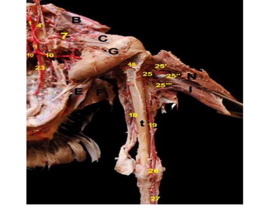
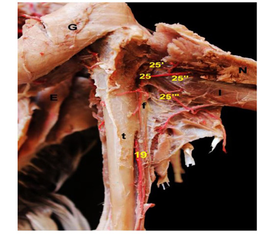
Figure 9:A photograph showing the branches and continuation of the fibular artery (Dorso-lateral view).
|
B. M.iliotibialis cranialis |
18. A.tibiais caudalis |
|
C. M.femorotibialis medialis |
19. A.tibialis cranialis |
|
G. M.gastrocnemius lateralis |
23. Pars synsacralis aortae |
|
E. M.flexor cruralis medialis |
25. A.fibularis |
|
I. M. tibialis cranialis |
25' R.ascendens; R.fibularis superficialis |
|
N. M. gastrocnemius pars intermedia |
25'' R.muscularis |
|
t. tibia |
25''' R.descendens; R.fibularis profundus |
|
1. Aorta descendens |
26. A.metatarsalis dorsalis communis |
|
4. A.pubica |
27. A.metatarsalis dorsalis medialis |
|
7. A.femoralis cranialis |
48. A. genicularis lateralis |
|
10. A.ischiadica |
|
the leg; the pars medialis, intermedia of Mm.gastrocnemis, popliteus, flexor halluces longus, flexor digiti longus and flexor perforates digiti III. It runs parallel to shaft of the tibia till reaches the caudal aspect of the hock joint where it ramifies into 3-4 fine branches.
A. tibialis cranialis
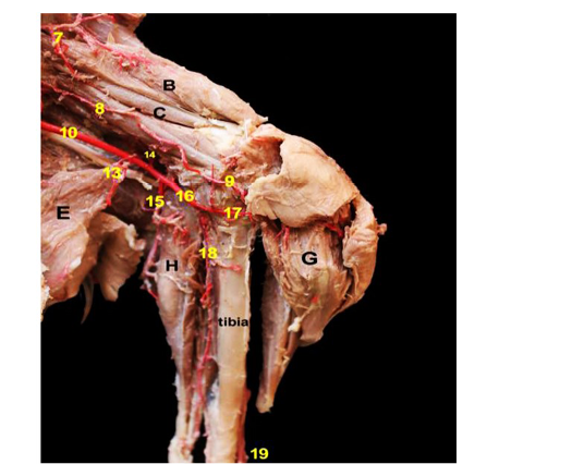
The cranial tibial artery (Figs.1/26 &2/34 & 4, 7,8, 9, 10/19); the direct continuation of the popliteal artery, passes caudo-ventrally to the lateral surface of the tibia between the M.popliteus and the proximal part of shaft of tibia. It descends into the caudal interosseous groove for about 0.3 cm to supply the Mm. flexor perforates digiti III and flexor digiti longus. It penetrates the interosseous membrane between tibia and fibula and transverses it to the cranial surface of tibia to supply Mm. tibialis cranialis and fibularis longus. The cranial tibial artery continues till reaches the hock joint and ramifies into 2-3 fine twigs. During its course, it detaches; the fibular and lateral tibial arteries and continues at the proximal end of the tarsometatarsus as common dorsal metatarsal artery.
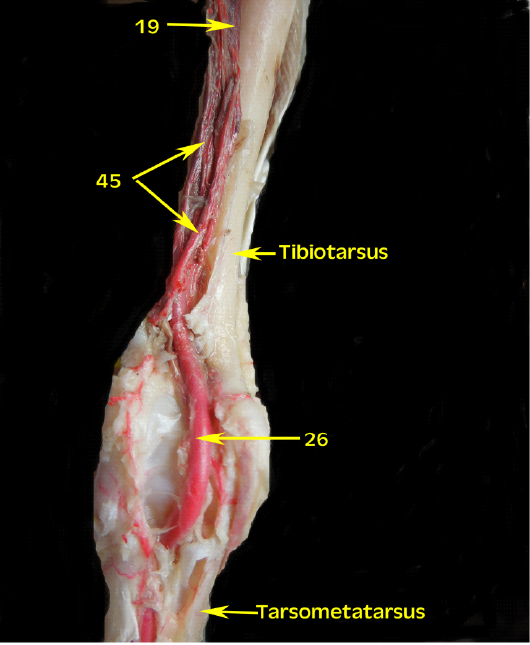
|
19. A tibialis cranialis |
|
26. A. metatarsalis dorsalis communis 45. Rete tibiotarsale |
A. fibularis
The fibular artery (Figs. 2/30 &8, 9/25); arises from cranio-lateral wall of the parent trunk. It passes laterally and pierces the proximal part of the interosseous foramen between tibia and fibula to deep face of M.tibialis cranialis and pars medialis of M.gastrocnemis where it bifurcates into; the descending and ascending branches. The former branch (Figs. 2/32 &8, 9/25’’’) is a small twig, distributed into the M. extensor digiti longus by fine radicles while the ascending branch (Figs. 2/31&8, 9/25’) is a fine short branch that passes cranio-dorsally between the head of fibula, proximal extremity of tibia and ramifies into 2-3 fine twigs to supply Mm.tibialis cranialis and fibularis longus. The fibular artery continues its course till give lateral geniculate artery (Fig.9/48) to supply lateral aspect of the stifle joint capsule.
A. tibialis lateralis
The lateral tibial artery is detached from the caudal aspect at the proximal third of the lateral surface of tibia. It courses through M.fibularis longus to reach the dorsal surface of the hock. It ramifies with the other muscular branches of cranial tibial artery in the extensor group of the leg.
The arterial blood supply of goose’s foot is supplied by two sets; the dorsal and planter.
The dorsal set:
The dorsal set involves of the Rete tibiotarsale and A. metatarsalis dorsalis communis
RETE TIBIOTARSALE
The rete tibio-tarsale (Figs. 2/35, 36 &10/45) is a network of multiple arteries and veins which is located on the distal third of the tibiotarsus and proximal part of tarsometatarsus. It comprises the collateral branches of the cranial tibial, deep fibular, superficial fibular and lateral sural arteriesthen continues distally to the dorsal aspect of tarsometatarsus as common dorsal metatarsal artery.
A. metatarsalis dorsalis communis
The common dorsal metatarsal artery (Figs. 2/39 &9,10,11/26); is the direct continuation of the cranial tibial artery at the flexor surface of tarsus and descends to dorsal surface of tarsometatarsus. Along its course; it gives off a perforating branch traverses the proximal vascular foramen to reach the plantar aspect forming the plantar set then continues on the dorsal surface of tarsometatarsus to be divided into medial and lateral dorsal metatarsal arteries.
A. metatarsalis dorsalis medialis
The medial dorsal metatarsal artery (Figs. 2/43 &9, 11/27); courses dorso-medially till reach the distal third of the tarsometatarsus where it gives off the medial digital I and medial digital II arteries. During its course, it detaches 2-3 fine arteries to supply Mm.extensor digitorum communis, extensor proprius digiti II and extensor halllucis longus.
A. metatarsalis dorsalis lateralis
The lateral dorsal metatarsal artery (Figs. 2/42 &11/28); descends on the tarso-metatarsus till reach the distal vascular foramen, through which it traverses to the planter side and participate in the formation of the planter arch. Along its course, it gives off fine branches to supply the tendon of the Mm. extensor digitorm communis, extensor proprius digiti II,extensor breves digiti III and IV.
II. The planter set:
The planter set of arteries is composed of the Aa.inter-metatarsales plantares and the planter arch.
1. Aa. Intermetatarsles Plantares Lateralis Et Medialis
The lateral and medial planter inter-metatarsal arteries (Fig.11/48); originate from the common dorsal metatarsal artery on the planter aspect of tarsus after its emergence from the proximal vascular foramen of tarso-metatarsus. Each bifurcates into an ascending and descending branches. The former one is denominated as the planter tarsal artery while the descending branch forms the planter metatarsal artery.
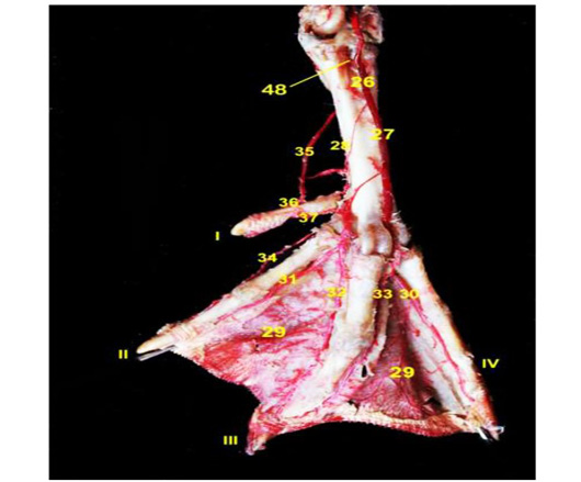
|
33. A.digitalis III lateralis |
26. A.metatarsalis dorsalis communis |
|
34. A.digitalis II medialis |
27. A.metatarsalis dorsalis medialis |
|
35.A. metatarsalis plantaris medialis |
28. A.metatarsalis dorsalis lateralis |
|
36. A.digitalis I medialis |
29. Rr. interdigitales |
|
37. A.digitalis I lateralis |
30. A.digitalis IV medialis |
|
48. Aa. intermetatarsales plantares |
31. A.digitalis II lateralis |
|
|
32. A.digitalis III medialis |
Aa. TARSALES PLANTARES
The plantar tarsal arteries (Figs. 2/37, 38 & 12/42), originated as short thin vessels from ascending branch of the planter inter-metatarsal artery. They ascend proximally on the planter sides of tarsus where they ramify on the tarsus and skin around the latter joint. Along their course; they joined the lateral sural arteries to supply the corresponding region.
Aa. METATARSALES PLANTARES LATERALIS ET MEDIALIS
The lateral and medial planter metatarsal arteries (Figs. 2/40, 41& 11,12/35& 12/41); the distal continuation of the descending branches of the planter inter- metatarsal artery. They proceed on the either sides of the planter aspect of tarso-metatarsus and nourish several muscles; Mm. flexor halluces longus, flexor digitorum longus, tendon of M.flexor digitorum profoundus and M.flexor digitorum superficialis by fine twigs. Finally, the planter metatarsal arteries unite with the lateral dorsal metatarsal artery to form the planter arch.
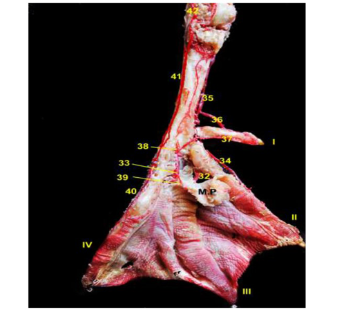
|
38. Arcus plantaris |
32. A.digitalis III medialis |
|
39. R. pulvinaris |
33. A.digitalis III lateralis |
|
40. A.digitalis IV lateralis |
34. A.digitalis II medialis |
|
41. A. metatarsalis plantaris lateralis |
35. A. metatarsalis plantaris medialis |
|
42. A. tarsalis plantaris |
36. A.digitalis I medialis |
|
M.P. metatarsal pad |
37. A.digitalis I lateralis |
Arcus Plantaris
The plantar arch (Figs. 2/44 &12/38) comprises the union of the medial, the lateral plantar metatarsal arteries by 1-2 fine transverse branches and the lateral dorsal metatarsal artery which pierces the distal vascular foramen of the tarsometatarsus. It is situated at the deep face of the common digital flexor tendon and runs distally till reaches the proximal extremity of the fetlock joint where it is divided into pulvinar branch and digital arteries.
R. PULVINARIS
The pulvinar branch (Fig. 12/39); arises from the planter arch in the majority of specimens. In few specimens, it emanates from a common stem with the lateral digital III, medial digital IV arteries. It descends to be distributed on the medial aspect of the metatarsal pad.
Aa. DIGITALES
The digital arteries (Fig.11/29); supply the digits from both medial and lateral sides. They arise from the plantar arch except the medial digital I (Figs. 2/45 &11,12/36), medial digitalII (Figs. 2/46 &11,12/34) and lateral digital IV (Figs.2/48 &12/40) arteries which are detached from medial dorsal metatarsal artery and lateral plantar metatarsal artery respectively. The lateral digital II (Figs. 2/46 &11/31) and the fine branch of medial digital III (Figs. 3/47 &11, 12/32) arteries are detached from lateral dorsal metatarsal artery. The digital arteries that spring from the plantar arch are; the lateral digital I artery (Figs. 2/45 &11,12/37), the lateral digital III artery (Figs.2/47 &11, 12/33) and the medial digital IV artery (Figs. 2/48 &11/30) which are erupted by a common stem from the plantar arch.
DISCUSSION
The chief arterial supply of the pelvic limb comprises the external iliac and ischiatic arteries, which is consistent with previous studies by (Havenga et al., 2020) in raptor, (El-Gammal, 2012) in chicken, (Can et al., 2010) in Japanese quail,(El-Nahla et al., 2010) in ostrich (Jianzhong et al., 2010) in chicken, (Dyce et al., 2002) in the avian species, (Kuru 1996, Baumel et al., 1993; King and Mclelland 1984, Nickel et al., 1977; Baumel, 1975, Mcleod et al.,1964; Nishida,1963) in domestic fowl. In contrast to (Bentley and Poople, 2009) in the Quail in the same respect, the main artery of the hind limb was the ischiatic artery which was released from the dorsal aorta.
The external iliac artery was a short trunk, arose from the lateral side of the descending aorta, within division between cranial and middle lobes of the kidney. This same observation was presented by (El-Nahla et al., 2010) in ostrich and (Kuru, 1996; Baumel et al.,1993, King et al., 1984; Baumel, 1975) in domestic fowl. (El-Gammal 2012, Dyce et al., 2002; Koch, 1973) in chicken and (El-Nahla et al., 2010) in ostrich reported that the external iliac artery arose from the descending aorta at the level of the knee joint, ventral to the body of synsacrum. However, the external iliac artery originated from the descending aorta at the level of the middle segment of the kidney (Havenga et al., 2020) in raptor, (Can et al., 2010) in Japanese quail, (Nickel et al., 1977) in domestic fowl and (Fitzgerald, 1969) in the European quail and (Evans, 1961) in the fowl.
The pubic artery is detached from the lateral aspect of the external iliac artery. This statement was asserted by (Koch, 1973, El-Gammal, 2012) in chicken, (El-Nahla et al., 2010) in ostrich and (Dyce et al., 2002, Kuru, 1996, Baumel et al., 1993, King et al., 1984, Baumel, 1975) in domestic fowl. (Havenga et al., 2020) in raptor observed that the external iliac artery gave off the femoral circumflex, gluteal and pelvic arteries. However, (Jianzhong et al., 2010) in chickens decided that the external iliac artery detached two branches; the femoral and circumflex femoral arteries that were distributed into the hind limb. On the other hand, (Nickel et al., 1977) had the opinion that the external iliac artery left the pelvic cavity where it divided into the pelvic and the femoral arteries in domestic fowl while (Can et al., 2010) in Japanese quail and (Fitzgerald, 1969) in the European quail mentioned that the external iliac artery emitted the umbilical (pubic) and the cranial gluteal arteries as well as the muscular branches that supplied the sartorius and the quadriceps femoris muscles. Moreover, (Baumel, 1975; Baumel et al., 1993) in domestic fowl stated that the umbilical artery was a large branch of the pubic artery which courses in the ventral extra-peritoneal fat to the umbilical scar of the abdominal wall.
The femoral artery represented the direct continuation of the external iliac artery and gave off the cranial coxal, the cranial femoral, and the medial femoral arteries. This statement was in accordance with (El-Gammal, 2012) in chicken, (El-Nahla et al., 2010) in ostrich, (Baumel et al., 1993; Baumel, 1975) in domestic fowls. On the other hand, (Jianzhong et al., 2010) claimed that the femoral artery gave off two branches; the acetabular branch and the lateral reticular artery in chicken, while (Can et al., 2010) in Japanese quail described that the femoral artery originated from the external iliac artery at the cranial aspect of the hip joint. However, (Nickel et al., 1977) in domestic fowl reported that the femoral artery arose from the external iliac artery and extended on the medial surface of the thigh to the flexor aspect of the knee where it became the a. genus suprema.
The ischiatic artery was the main source of the blood supply of the pelvic limb of goose, such statement was similar to that asserted in chicken (El-Gammal, 2012), Japanese quail (Can et al., 2010), ostrich (El-Nahla et al., 2010), rooster, drake and pigeon (Kurtul and Haziroglu 2002), and domestic fowls (Kuru 1996, Baumel et al., 1993; King et al., 1984). The ischiatic artery originated from ventro-lateral aspect of the abdominal aorta, caudal to the origin of the external iliac artery, in contrast with that by (Havenga et al., 2020) in raptor. The ischiatic artery emerged from descending aorta at the middle axis of the division renalis media (Havenga et al., 2020), while in chicken (El-Gammal, 2012), Japanese quail (Can et al., 2010), and domestic fowls (Baumel et al., 1993; Baumel 1975) the ischiatic artery arose from lateral aspect of the aorta caudal to the origin of the femoral artery. On the other hand,the ischiatic artery detached from the aorta at the junction of the middle and caudal renal divisions, caudal to the origin of the external iliac artery in ostrich (El-Nahla et al., 2010) while in domestic fowl showed that the ischiatic artery sprang from the aorta at the level of the hip joint (Nickel et al., 1977).
The extra pelvic distribution of the ischiatic artery, the current work demonstrated that it gave off the caudal coxal artery after leaving the pelvis through ilioischiatic foramen. These findings denied that had been mentioned by (El-Gammal, 2012) in chicken, (Baumel et al., 1993, Nickel et al., 1977; Baumel 1975) in domestic fowl.
The popliteal artery was the direct continuation of the ischiatic artery in the leg region. The same result was mentioned by (El-Gammal, 2012) in chicken, (Can et al., 2010) in Japanese quail, (El-Nahla et al., 2010) in ostrich, (Nickel et al., 1977, Baumel et al., 1993, Baumel 1975, Fitzgerald 1969; Mcleod et al., 1964) in domestic fowl. The popliteal artery in goose gave off; the proximal geniculate, medial tibial and caudal tibial arteries; similar information was given by (El-Gammal, 2012) in chicken. On the other hand, (El-Nahla et al., 2010) in ostrich, (Baumel et al., 1993, Baumel, 1975, Fitzgerald, 1969; Mcleod et al., 1964) in domestic fowl stated that the popliteal artery gave off the caudal tibial artery. (Can et al., 2010) explained that the popliteal artery divided into cranial tibial and caudal tibial arteries in the caudo-medial aspect of the knee joint in Japanese quail.
The popliteal artery continued as the cranial tibial artery. This result was confirmed previously in chicken (El-Gammal, 2012), in ostrich (El-Nahla et al., 2010), in domestic fowl (Baumel et al., 1993, Baumel 1975, Fitzgerald, 1969; Mcleod et al., 1964), while it was contrary to that reported in domestic fowl as the popliteal artery terminated into cranial and lateral tibial arteries (Nickel et al., 1977).
The cranial tibial artery was considered the direct continuation of the popliteal artery. This statement was in accordance with (Tolba and Daghash, 2014) in goose, (Farag, 2013) in duck, (El-Gammal, 2012) in chicken, (EL-Nahla et al., 2010) in ostrich, (Baumel et al., 1993; Midtgard, 1982) in the different birds, (Nickel et al., 1977, Baumel, 1975; Mcleod et al., 1964) in domestic fowl. It gave off the fibular and lateral tibial arteries, which is a similar finding to that presented by (El-Gammal, 2012) in chicken.
The cranial tibial artery was continued as common dorsal metatarsal artery at the proximal end of the tarsometatarsus; simulated that had been mentioned by (Tolba and Daghash, 2014) in goose, (Farag, 2013) in duck, (El-Gammal, 2012) in chicken, (El-Nahla et al., 2010) in ostrich, (Baumel et al., 1993, Nickel et al., 1977, Baumel, 1975; Mcleod et al., 1964) in domestic fowl.
The rete tibio-tarsalewas a network of multiple arteries and veins which located on the distal third of the tibiotarsus and proximal part of tarsometatarsus; in agreement with (Tolba and Daghash, 2014) in goose, (Farag, 2013) in duck, (Dursun 2002) in the domestic birds, (Midtgard 1981) in the different birds and (Fitzgerald, 1969) in the coturnix quail. On the other hand, (El-Gammal, 2012) in chicken and (Baumel, 1975) in domestic fowl revealed that the common dorsal metatasrsal artery with collateral branches of the cranial tibial artery formed the multiple vessels referred to as the rete tibialis dorsalis while (Can et al., 2010) in Japanese quail recorded that the rete tibio-tarsale was a net made up of the branches of the cranial tibial artery on the cranial aspect of the tibia.
The common dorsal metatarsal artery continued on the dorsal side of tarsometatarsus to divide into the medial and the lateral dorsal metatarsal arteries which was a similar observation to that reported in goose (Tolba and Daghash, 2014),in duck (Farag, 2013), in chicken (El-Gammal 2012) and in domestic fowl (Nickel et al., 1977).
Concerning the plantar tarsal arteries, they originated from ascending branch of the planter inter-metatarsal artery, in agreement with (Tolba and Daghash, 2014) in goose and (Farag, 2013) in duck; disagreed with (El-Gammal, 2012) in the chicken and (Can et al., 2010) in the Japanese quail who revealed that the rete tarsi-dorsalis gave off two branches; the lateral and the medial planter tarsal arteries, while (El-Nahla et al., 2010) in the ostrich, (Baumel et al., 1993, Nickel et al., 1977; Baumel 1975) in the domestic fowl reported that the medial and the lateral planter tarsal arteries arose from the common dorsal metatarsal artery on the dorsal aspect of the tarsometatarsus.
The plantar arch comprised the union of the medial, the lateral plantar metatarsal arteries and the lateral dorsal metatarsal artery, similar to that reported by (Tolba and Daghash, 2014) in goose and (Farag, 2013) in duck. In contrast, the descending branches of the medial and lateral planter tarsal artery were connected inside the metatarsal pad with the lateral and medial dorsal metatarsal arteries to form the Arcus plantaris which was responsible for detaching the digital arteries in the sole of the foot in chicken (El-Gammal, 2012). The pulvinar branch arose from the planter arch in the majority of the cases while in few specimens was emanated from a common stem with the lateral digital III, medial digital IV arteries (Tolba and Daghash, 2014) in goose and (Farag, 2013) in duck. Moreover, the pulvinar arteries arose from the common dorsal metatarsal artery at the level of the meta-tarso-phalangeal joint (El-Nahla et al., 2010) in ostrich, (Baumel et al., 1993, Nickel et al., 1977; Baumel, 1975) in domestic fowl.
In contrast to the present study, the dorsal metatarsal artery gave off the digital arteries in goose (Waxman et al., 2015), while all digital arteries erupted from the plantar arch (Tolba and Daghash, 2014) in goose and (Farag, 2013) in ducks. (Tolba and Daghash, 2014) added that the lateral digital II artery and lateral digital IV originated from the medial dorsal metatarsal artery and lateral plantar metatarsal artery respectively, while the arteries of the 2nd digit and lateral IV digital artery erupted from medial dorsal metatarsal artery and lateral planter metatarsal artery, respectively (Farag, 2013). However, the digital arteries of the 1st digit, medial II digital artery, medial III digital artery and lateral IV digital artery originated from the planter arterial arch (El-Gammal, 2012) in chicken and the lateral III digital artery and medial IV digital artery arose from lateral dorsal metatarsal artery (Can et al., 2010) in Japanese quail.
CONCLUSION
Arterial blood supply of the pelvic limb of the geese was mainly obtained through the external iliac and ischiatic arteries. The external iliac artery only supplies the pelvic limb of the geese to the level of the stifle region, while the ischiatic artery is responsible for supplying the entire limb, changing its name according to the region of the limb it supplies. The ischiatic artery terminates after giving off the sural artery and continues in the leg region as popliteal artery. The popliteal artery supplies the knee and leg regions, whereas the cranial tibial artery, the continuation of the popliteal artery, is responsible for supplying the foot with its own branches. The Rete tibiotarsale is playing a role in thermoregulation of the leg temperature through heat conservation or dissipation due to the presence of extensive arteriovenous anastomosis.
AUTHOR CONTRIBUTIONS
All authors contributed to the experimental design, sampling, and reviewing the scientific content. Gamal A. Swielim, Khaled Abo‐EL‐Sooud and Samar M. EL‐Gammal performed the scientific and grammatical revisions to the manuscript. Meray N. Ramsis performed the experiments, data collection and writing the paper.
CONFILCT OF INTErEsT
The authors have declared no conflict of interest.
REFERENCES


