Advances in Animal and Veterinary Sciences
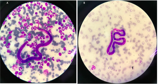
Microfilariae in the cat (A) and the dog (B).
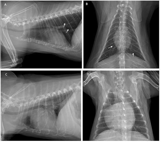
Thoracic radiograph shows the opaque rings of a bronchial pulmonary lung pattern (white arrow) in the cat (A and B), and enlarged heart of the dog (C and D) with heartworm infection.
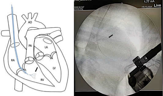
Mini vascular snare device (black arrow) with working space 4-8 millimeter was inserted into the right cardiac chamber with fluoroscopic guidance.
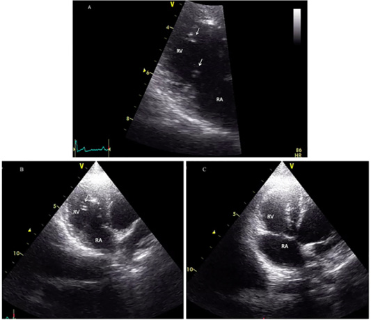
Echocardiographic image showing heartworms (white arrow) in the right ventricle of the infected cat (A and B) and after adult heartworm removal in C. Before the surgical intervention (A and B), heartworms (white arrow) are visible in the right ventricle (RV), no heartworm is visible after the procedure (C).
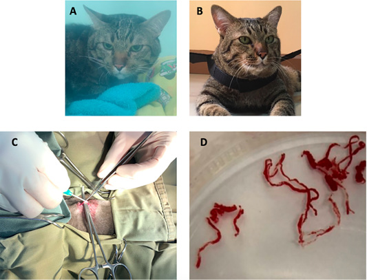
The image showing a cat before surgical intervention (A) in the oxygen chamber, and after adult heartworm removal in (B). The image during the surgical procedure (C), adult heartworms (D) are removed from the right ventricle of the heart.


