Advances in Animal and Veterinary Sciences
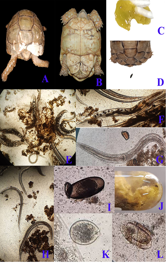
A & B; Clinical presentation of gastroenteritis in Egyptian tortoise, photographs showing multiple areas of white discoloration, pores and cracks in carapace and plastron respectively which indicates severe minerals deficiency due to diarrhea. C: photograph showing diarrhea with the presence of numerous motile worms. D: photograph showing the normal configuration of stool. Fecal samples analysis of diarrheic tortoises; E: Severe infestation with large numbers of adult males, females and larvae of Oxyurid species (4X). F: Adult female Oxyurid species contains large number of eggs (10X). G: Adult male Oxyurid species (10X). H: Larvae of Oxyurid species (4X). I: Oxyurid species egg showing central embryo (40X). J: Gross appearance of diarrheic stool with adult pin worms (Oxyurid species). K: Plantadium species trophozoite (40X). L: Nyctotheris species trophozoite (40X).
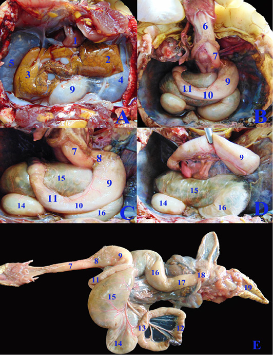
A, B, C, D; The coelomic cavity of Healthy female Desert Egyptian tortoise after removing the plastron (fresh specimen). E: Photograph showing separated healthy GIT. 1. Heart 2. Left lobe of liver 3. Right lobe of liver 4. Left lung 5. Right lung 6. Trachea 7. Esophagus 8. Gastro esophageal sphincter 9. Stomach 10. Pyloric sphincter 11. Duodenum 12. Jejunal loops. 13. Ileum 14. Cecum 15. Ascending colon 16. Transverse colon 17. Descending colon 18. Cloaca.
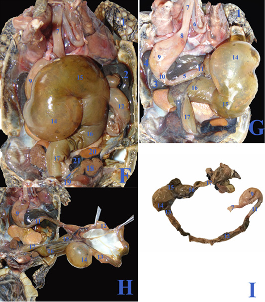
F, G, H; The coelomic cavity of diseased male Desert Egyptian tortoise after removing the carapace (the tortoise was dissected after death freshly). I: Photograph showing separated diseased GIT. 1. Right forelimb 2. Right lobe of liver 3. Left lobe of liver 4. Spleen 5.Pancreas 6. Right bronchus 7. Esophagus 8. Gastro esophageal sphincter 9. Stomach 10. Pyloric sphincter 11. Duodenum 12. Jejunal loops. 13. Ileum 14. Cecum 15. Ascending colon 16. Transverse colon 17. Descending colon 18. Right kidney 19. Right adrenal gland 20. Right testes 21. Right epididymis.
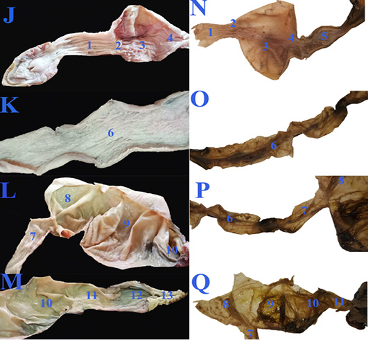
The macroscopic internal structure of the healthy GIT (J, K, L &M) and the diseased GIT (N, O, P &Q) of Desert Egyptian tortoise. 1. Esophageal mucosa 2. Gastro esophageal sphincter mucosa 3. Gastric mucosa 4. Pyloric sphincter mucosa 5. Duodenal mucosa 6. Jejunal mucosa 7. Ileum mucosa 8. Cecal mucosa 9. Ascending colon mucosa 10.transverse colon mucosa 11. Descending colon mucosa 12. Cloaca 13. Vent.
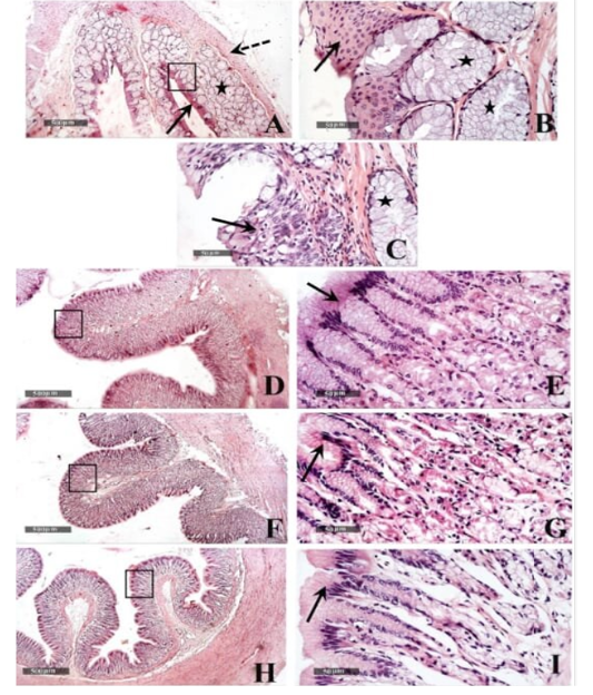
Histological features of esophageal samples (A, B & C), cardiac region and glands (D & E), fundic region and glands (F& G); pyloric region and glands (H & I). H&E stain – 40X & 400 X magnifications.
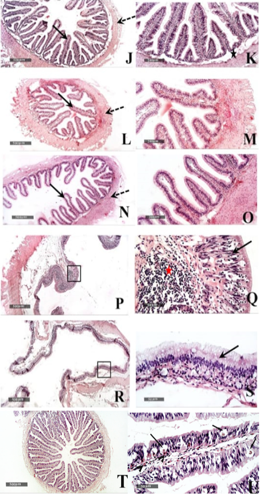
Histological features of Duodenum (J & K), Jejunum (L & M), and Ileum (N &O) - 40 X & 200 X magnifications. Cecum (P & Q) and colon (R & S) H&E stain – 40X & 400 X magnifications. T&U: showing Microscopic examination of intestinal/duodenal samples revealed many intra epithelial parasitic stages all over intestinal villi (arrow) accompanied with underlying lamina propria or intra epithelial eosinophilic infiltrates in (arrowhead).


