Advances in Animal and Veterinary Sciences
Research Article
Adv. Anim. Vet. Sci. 9(3): 446-452
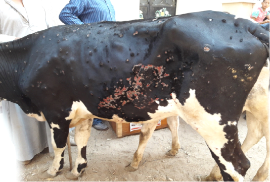
Figure 1
Multiple intradermal skin nodules with sloughing of many of them leaving deep ulcers due to infection with LSDV
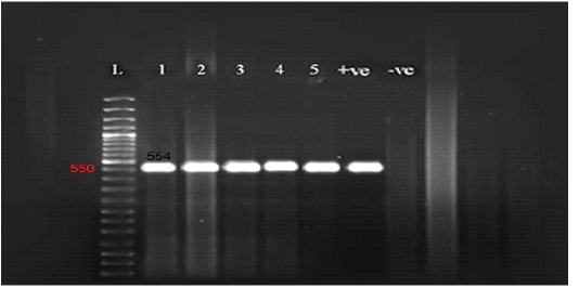
Figure 2
Gel electrophoresis of amplified partial GPCR gene
Lane L (50 bp Ladder marker), lanes 1-5 (positive samples), lane +ve (control positive), and lane –ve (control negative).
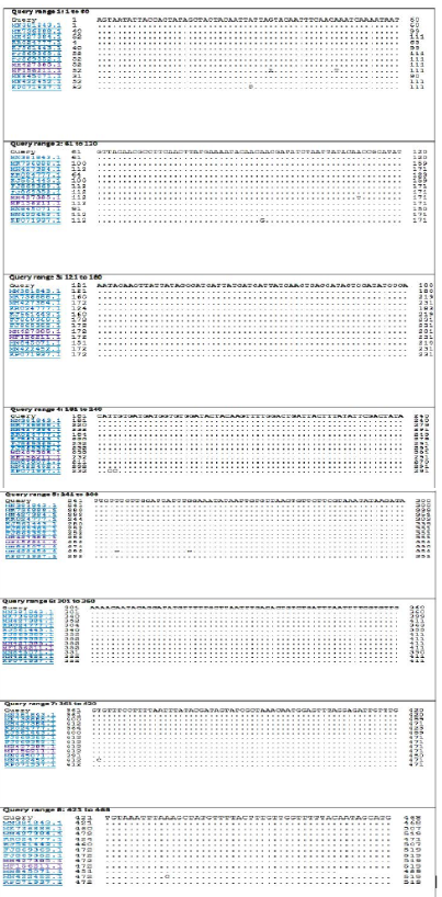
Figure 3
Nucleotide sequence alignment identity of partial GPCR gene of the present LSDV outbreak isolate with different LSDV field isolates.
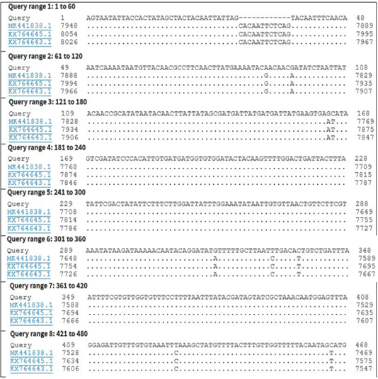
Figure 4
Nucleotide sequence alignment identity of partial GPCR gene of the present LSDV outbreak isolate with different LSDV vaccinal isolates.
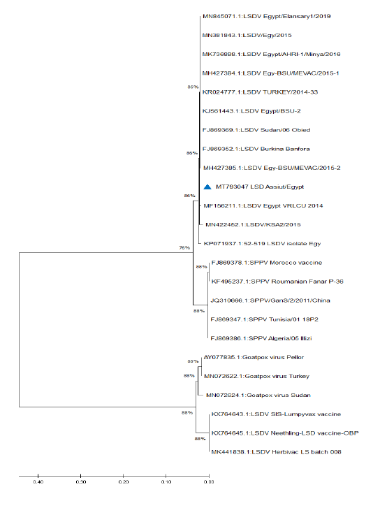
Figure 5
Phylogenetic analysis of different capripoxviruses based upon the nucleotide sequences of GPCR gene.


