Advances in Animal and Veterinary Sciences
Short Communication

Ocular Biometry and Ophthalmic Parameters of Normal Eyes in French Bulldog Healthy Dogs
Talita Franco Andrade1, Luíza Rodrigues Dale Vedove Moreno2, Felipe Franco Nascimento3, João Victor Goulart Consoni Passareli3, Vinícius dos Santos Rosa4, Thais Caroline da Silva Santos4, Rejane Batista Brinholi5, Silvia Franco Andrade6*
1Resident of Image of the Veterinary Hospital of Faculty of Veterinary Medicine, Unesp, Araçatuba, SP, Brazil; 2Resident of Image of Veterinary Hospital of Faculty of Veterinary Medicine, University of Oeste Paulista (UNOESTE), Presidente Prudente, SP, Brazil; 3Post Graduate Program in Animal Science, University of Oeste Paulista (UNOESTE); 4Faculty of Veterinary Medicine, UNOESTE; 5Department of Image of Veterinary Hospital of UNOESTE; 6Department of Ophthalmology of Veterinary Hospital of UNOESTE.
Abstract | The objective was to perform ocular biometry and ophthalmic examination in dogs of the French Bulldog breed. We evaluated 72 normal eyes of 36 healthy French Bulldog dogs, 20 males and 16 females, aged from 1 to 7 years. Ocular ultrasound was performed in B mode with a 10 MHz linear probe, and the following parameters of ocular biometry were evaluated: horizontal axial diameter (HAD) of the eye, lens thickness (LT), anterior chamber depth (ACD), and vitreous chamber depth (VCD). The ophthalmic exams performed were as follows: Schirmer Tear Test-1 (STT-1), rebound tonometry (Tonovet), Tear Film Break-up Time (TBUT), and Fluorescein Test (FT). The ocular biometry results (mean±SD) in millimeters were: 20.5 ± 0.6 (HAD), 3.8 ± 0.4 (ACL), 7.4 ± 0.3 (LT) and 9.4 ± 0.4 (VCL), and from the ophthalmic exams (mean±SD) were: 24 ± 3.7 (16.5-33.5) mm/min (STT-1), 20.0 ± 3.7 (13.5-27.0) mmHg (rebound tonometry), and 24.4 ± 6.3 (16-40) seconds (TBUT), and FT was negative in all dogs. The obtained results can serve as parameters of normal values of ocular biometry and ophthalmic exams for ambulatory routines, intraocular surgeries, and future ophthalmic studies in this breed.
Keywords | Ocular ultrasound, Ocular biometry, Ophthalmic exams, French Bulldog, Dogs
Received | Septeber 30, 2020; Accepted | December 04, 2020; Published | January 15, 2021
*Correspondence | Silvia Franco Andrade, Department of Ophthalmology of Veterinary Hospital of UNOESTE, Rodovia Raposo Tavares Km 572, Bairro Limoeiro, Presidente Prudente, SP, Brazil, CEP 19067-175; Email: silviafranco@unoeste.br
Citation | Andrade TF, Moreno LRDV, Nascimento FF, Passareli JVGC, Rosa VdS, Santos TCdS, Brinholi RB, Andrade SF (2020). Ocular biometry and ophthalmic parameters of normal eyes in french bulldog healthy dogs. Adv. Anim. Vet. Sci. 9(3): 438-441.
DOI | http://dx.doi.org/10.17582/journal.aavs/2021/9.3.438.441
ISSN (Online) | 2307-8316; ISSN (Print) | 2309-3331
Copyright © 2021 Andrade et al. This is an open access article distributed under the Creative Commons Attribution License, which permits unrestricted use, distribution, and reproduction in any medium, provided the original work is properly cited.
INTRODUCTION
Ocular ultrasound (US) is a non-invasive, painless diagnostic method and can be considered relatively inexpensive, being a relevant diagnostic tool in the veterinary routine of small animals. The clinical indications for ocular US include ocular traumatic alterations, foreign body investigation, intraocular hemorrhage, lens dislocation, retinal detachment, and any opacity that makes the ophthalmoscope exam impossible. Intraocular and retrobulbar tissues and masses are visualized, evaluated sonographically and can be characterized by ultrasound-guided aspiration biopsy (Cottrill et al., 1989; Gaidon et al., 1991; Boroffka et al., 2006). In addition, it is an important tool for performing ocular biometrics for globe measurements which can be used to determine intraocular or intraorbital prosthetic size. These measurements are also helpful in calculating appropriate intraocular lens (Dietrich, 2013; Toni et al., 2013; Boillot et al., 2014; Kobashigawa et al., 2015).
The objective of this study was to perform ocular biometry and specific ophthalmologic diagnostics in healthy dogs of the French Bulldog breed, an brachycephalic dog, that are prone to eye disease, and currently a very popular breed worldwide, since these important ocular parameters for this breed do not exist in the literatures and can serve as parameters of normal values of ocular biometry and ophthalmologic examination.
The study was approved by the Ethics Committee on Animal Use (CEUA) of UNOESTE (Protocol nº.3853) and was carried out according to the rules of ARVO (Association for Research in Vision and Ophthalmology - Statement for the use of animals in ophthalmic and visual research). We evaluated 72 normal eyes of 36 healthy French Bulldog dogs, attended to at the UNOESTE Veterinary Hospital, Presidente Prudente, SP, Brazil. There were 20 males and 16 females, mean weight was 12.2±1.5 kg, and the aged range was from 1 to 7 years.
The inclusion criteria for French Bulldog dogs in the research was the normality in the parameters of the clinical exams (temperature, heart rate and respiratory rate and blood pressure), laboratory tests (blood count, urea, creatinine, alanine aminotransferase and alkaline phosphatase), clinical ophthalmic signs as identified using a portable slit lamp (SL-15, Kowa, Japan) and ophthalmic tests (Direct and consensual pupillary reflex test (DPRT and CPRT), Schirmer Tear Test-1 (STT-1) (Laboratory Ophthalmos, Brazil), rebound tonometry (Tonovet®, Finland), Tear Film Break-up Time (TBUT), and Fluorescein Test (FT) (Laboratory Ophthalmos, Brazil).
To obtain the data, ocular biometry was performed by ocular ultrasound (Mindray ultrasound device, model Z6 Vet, Shenzhen, China) in B-mode with the 10 MHz linear probe (Mindray linear probe model 7L4P, Shenzhen, China). Dogs were delicately contained sitting or in prone position. Blepharostase was manual, and the evaluation was performed under low light conditions after instillation of 1 drop of anesthetic eye drops (0.5% proxymetacaine, Anestalcon®, Allergan, São Paulo, Brazil). Sterile gel was used as a means of contact, using the transcorneal or transpalpebral method (Figure 1). The following parameters were evaluated: horizontal axial diameter (HAD) of the eye, anterior chamber depth (ACD), lens thickness (LT), and vitreous chamber depth (VCD).
The values of ocular biometrics and ophthalmic tests were expressed as the mean ± SD, and 95% confidence interval. The ocular biometry results (mean±SD) in millimeters were as follows: 20.5 ± 0.6 (HAD), 3.8 ± 0.4 (ACD), 7.4 ± 0.3 (LT), and 9.4 ± 0.4 (VCD). The B-scan image of a female French Bulldog is in Figure 2. The results from the ophthalmic exams (mean±SD) were as follows: 24 ± 3.7 (16.5-33.5) mm/min (STT-1), 20.0 ± 3.7 (13.5-27.0) mmHg (rebound tonometry), and 24.4 ± 6.3 (16-40) seconds (TBUT). In all dogs, the DPRT and CPRT were positive and the FT negative.
Several studies of ocular biometry have been described in horses (McMullen and Gilger, 2006), cats (Gilger et al., 1998), chinchillas (Lima et al., 2010), bald eagles (Khun et al., 2015), and dogs, from different breeds, such as Beagles (Boroffka et al., 2006), Shih Tzus (Kobashigawa et al., 2015), Eurasier dogs (Boillot et al., 2014), brachycephalic dogs (Toni et al., 2013) and mesocephalic and dolichocephalic dogs (Cottrill et al., 1989; Chiwitt et al., 2017). The data obtained in the present study of ocular biometrics in 36 French Bulldogs are the first study of ocular biometrics specific to this breed, and are similar to those described in a study published in 10 brachycephalic dogs (Toni et al., 2013), and by another study with different breeds, among them some of small size like the French Bulldogs, as in 10 Cavalier King Charles Spaniel, 6 Border Terrier, 8 Jack Russel Terrier and 8 West Highland White Terrier (Chiwitt et al., 2017). The values obtained from ophthalmic exams, STT-1, rebound tonometry and TBUT are within normal values for the canine species (Featherstone and Heinrich, 2013).
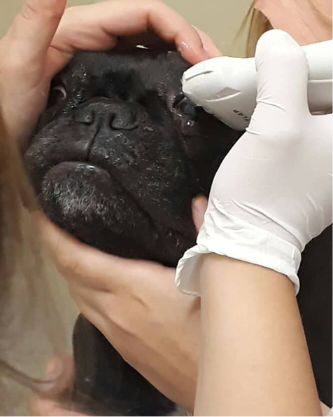
Figure 1: Ocular US evaluation in a female French Bulldog. Delicately contained in sitting position. Blepharostase was manual, and the exam was performed after instillation of 1 drop of anesthetic eye drops and sterile gel was used as a means of contact, using the transcorneal method.
In summary, the results obtained in French Bulldogs can serve as parameters of normal values of ocular biometry and ophthalmic exams for the ambulatory routine, intraocular surgeries, and future ophthalmic studies in this breed.
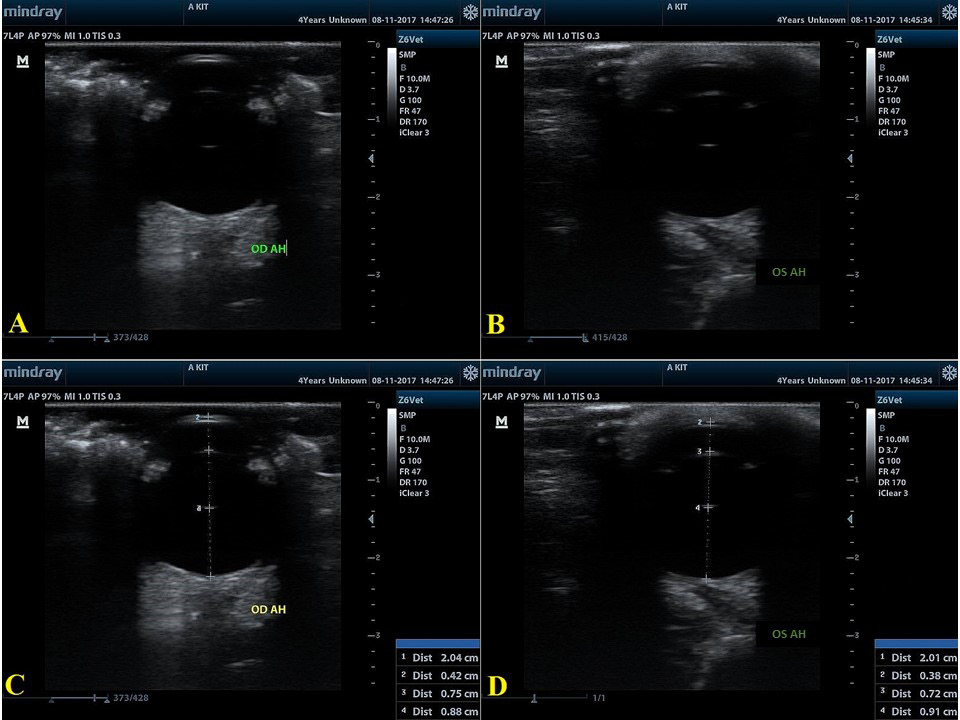
Figure 2: Representative B-scan ultrasonogram of female French Bulldog (A) and (B) representative B-scan ultrasonogram after optimal positioning was achieved and in (C) and (D) after the measures were done. Measurement values: Right eye (OD): (C) HAD = 2.04 cm (20.4 mm); ACD= 0.42 cm (4.2 mm); LT= 0.75 cm (7.5 mm); VCD= 0.88 cm (8.8 mm). Left eye (OS): (D) HAD= 2.01 cm (20.1 mm); ACD= 0.38 cm (3.8 mm); Lens Thickness= 0.72 cm (7.2 mm); VCD= 0.91 cm (9.1 mm).
Acknowledgments
The authors would like to thank the Postgraduate Program of UNOESTE for financial support.
Author’s Contribution
Talita Franco Andrade, Luíza Rodrigues Dale Vedove and Rejane Batista Brinholi were responsible for ocular ultrasound exams. Vinícius dos Santos Rosa and Thais Caroline da Silva were responsible for clinical and laboratory screening exams. Felipe Franco Nascimento, João Victor Goulart Consoni Passareli and Silvia Franco Andrade were responsible for ophthalmic examinations.
Conflict of interest
The authors have declared no conflict of interest.
References


