Advances in Animal and Veterinary Sciences
Case Report
Adv. Anim. Vet. Sci. 9(2): 315-319
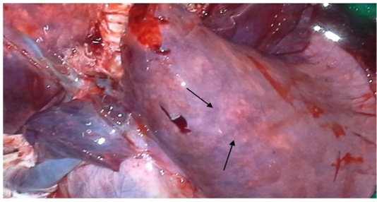
Figure 1
Congested and consolidated pneumonic lung with dark red discoloration (arrow)
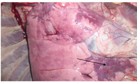
Figure 2
Congestion and consolidation of cardiac lobe of lung (Arrow)
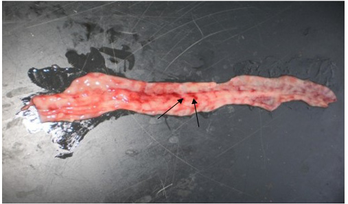
Figure 3
Zebra striping in colon
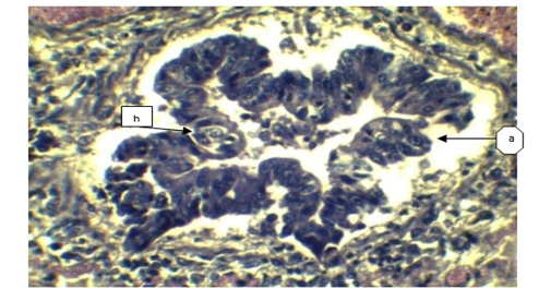
Figure 4
a) Sloughing off surface epithelium of bronchi; b) Necrosis of bronchiolar epithelium. (H&E x 40).
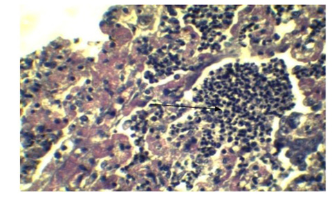
Figure 5
Infiltration of inflammatory cell in Lung parenchyma (Arrow), feature of interstitial pneumonia. (H&E x 40)
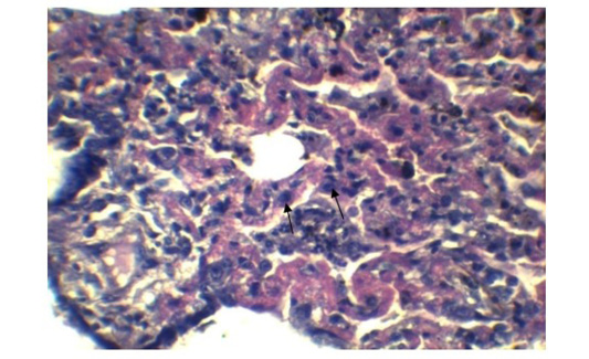
Figure 6
Expanded alveolar wall due to prominent type-II pneumocytes. (H&E x 40).
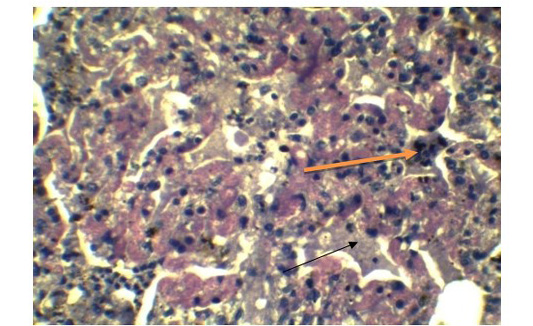
Figure 7
Presence of syncytial cell in the alveoli (Yellow arrow). Presence of fibrin exudates in parenchyma (Black arrow) (H&E x 40).

Figure 8
Inflammatory cells in the submucosa (Arrows). (H&E x 10).
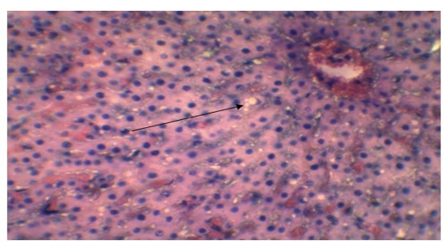
Figure 9
Vacuolar or fatty degeneration in the cytoplasm of hepatocyte (Arrow). (H&E x 40).


