Advances in Animal and Veterinary Sciences
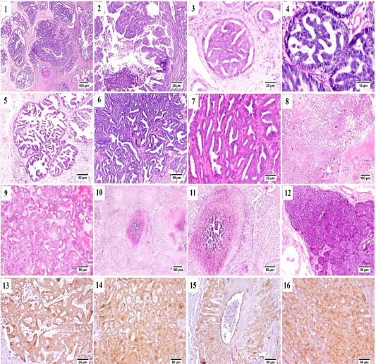
Mammary gland of cat , H&E stained, showing (1) papillary carcinoma, (2) Micropapillary carcinoma, (3) Ductal carcinoma in situ, (4) higher magnification, frequent mitosis of intraductal neoplastic cells, (5) cystic papillary carcinoma, (6) Tubular carcinoma, (7) higher magnification, tubular carcinoma, (8) cribriform carcinoma, (9) higher magnification, cribriform carcinoma, (10) comedocarcinoma, (11) higher magnification, comedocarcinoma with central necrotic area, (12) Solid carcinoma, (13) – (16) Positive Immunostaining of CK 8/18 in the different histologic patterns of carcinoma.
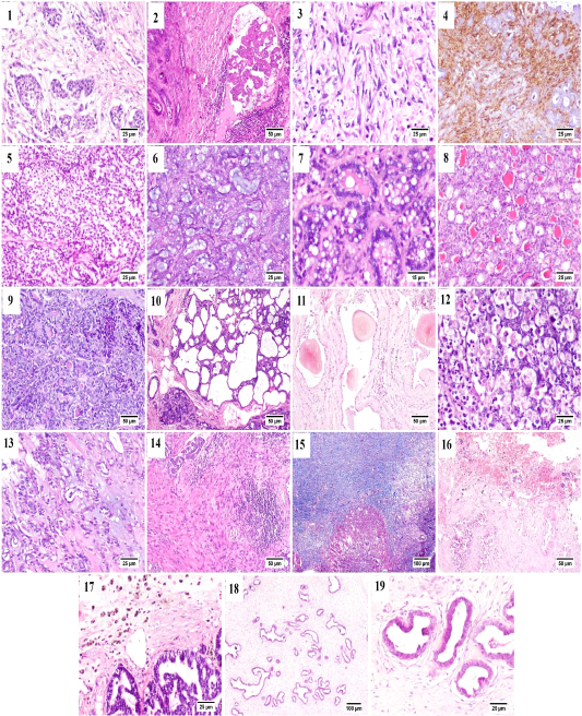
Mammary gland of cat, showing (1) Desmoplasia, (2) vascular invasion by anaplastic cells, (3) Pleomorphic population of myoepithelial cells, (4) Vimentin positive cells (immunostaining), (5) Glycogen-rich carcinoma, (6) mucinous carcinoma, (7) lipid-rich carcinoma, (8) lobular hyperplasia, (9) lobular hyperplasia with atypia, (10) cystically dilated ductules, (11) Duct ectasia with corpora amylacea, (12) Apoptosis with karyorrhexis, (13) Myxomatous change, (14) Stromal fibroplasia with lymphocytic infiltration, (15) proliferating fibrous tissue stained blue with MTC stain, (16) Extensive hemorrhage, (17) hemosiderin-laden macrophages, (18) fibroepithelial hyperplasia and (19) higher magnification, proliferating ductal epithelial tissue with surrounding myoepithelial cells and fibrous stroma.
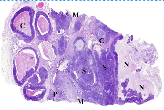
WSI of feline mammary gland tumor, resized, illustrating (C) Comedocarcinoma, (P) Tubulopapillary carcinoma, (S) Solid carcinoma, (N) necrosis and (M) surgical margins.


