Advances in Animal and Veterinary Sciences
Case Report
Adv. Anim. Vet. Sci. 9(1): 21-25
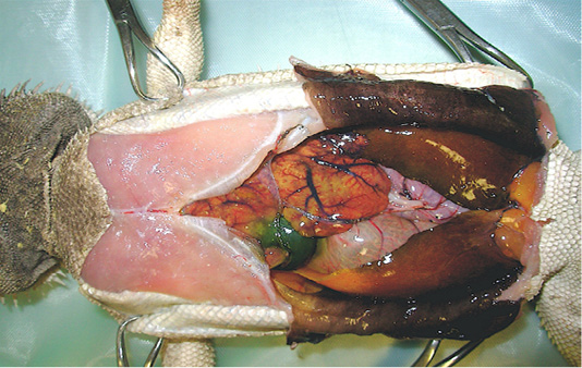
Figure 1
Necropsy examination of bearded dragon showing a moderate amount of sero-sanguineous fluid in abdominal cavity; a slightly enlarged liver with pale yellow appearance; a moderately enlarged and discoloured kidneys; and congested and hyperaemic lungs.
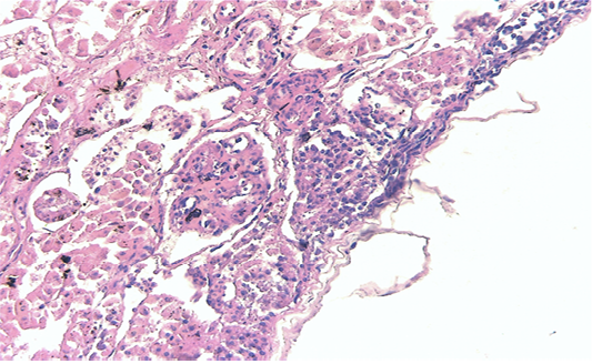
Figure 2
Kidney showing large multifocal areas of coagulative necrosis, associated with marked marginal hyperemia, interstitial nephritis, with presence of mononuclear and polymorphonuclear cells (H & E × 40, bar 100 μm).
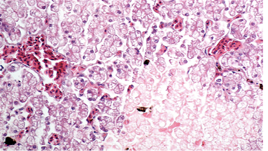
Figure 3
Liver showing cellular swelling and diffuse microvacuolar fatty change of hepatocytes, associated with scattered groups of necrotic hepatocytes; sinusoids were enlarged and congested (H & E × 20, bar 100 μm).
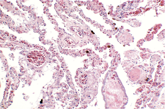
Figure 4
Lungs showing areas of extensive hyperaemia (H & E × 10, bar 100 μm).


