Advances in Animal and Veterinary Sciences
Research Article

Topographical and Morphological Studies on the African Helmeted Turtle (African Side-Necked Turtles - Pelomedusa Subrufa) with Special Reference to its Coelomic Cavity
Yara S. Abouelela*, Reem R. T.
Department of Anatomy and Embryology, Faculty of Veterinary Medicine, Cairo University, Giza 12211, Egypt.
Abstract | African Side-necked Turtle is a popular pet, used as food in addition to its ecological importance. It lives in freshwater at Sub-Saharan Africa, and in southern Yemen, of family Pelomedusidae which has unusual movements of the head. Five female juvenile African helmeted turtles were anesthetized with Xylazine 1mg/Kg/IM and ketamine 20mg/Kg/IM, after that, they were sacrificed by sodium thiopental at 2.5% in the lethal doses of 60mg/Kg/IV, then opened at the inguinal scute and dissected freshly. The absence the diaphragm leads to nonattendance of the pleural and abdominal cavities which substituted by the presence of ventral coelomic area and dorsal retro-coelomic area divided by the horizontal septum (transverse pleuroperitoneal membrane). Concerning the digestive tract and associated organs; the openings of the pharynx were six openings; paired internal choanal opening and Eustachian tubes openings, single glottis and aditus esophagus. The diameters and length of the small intestine parts differs. The jejunum was long extending from the right coelom to the left coelom. A small cecal bulb was present at the right of the median plane. The liver has five lobes; right medial and lateral, left medial and lateral and middle lobes, also a gall bladder was present. The compact pancreas has head, long body and triangular tail. In the respiratory organs; the lungs were very long covering more than half of the carapace’s length till reached caudally to ovary and the kidneys. Both kidneys were elliptical located at retro coelomic area at the same level in the pelvis region in relation to adrenal gland, ovary, and lung. The juvenile female reproductive system of the turtle consisted of a pair of ovaries, two oviducts, mesovarium and a mesosalpinx. The paired immature Ovaries were elliptical elongated granulated in appearance with yellow coloration.
Keywords | River turtle, Coelomic cavity, Anatomy, Digestive system, Septum horizontale
Received | June 13, 2020; Accepted | August 15, 2020; Published | November 15, 2020
*Correspondence | Yara S Abouelela, Department of Anatomy and Embryology, Faculty of Veterinary Medicine, Cairo University, Giza 12211, Egypt; Email: yarasayed89@gmail.com
Citation | Abouelela YS, Reem RT (2020). Topographical and morphological studies on the african helmeted turtle (african side-necked turtles - pelomedusa subrufa) with special reference to its coelomic cavity. Adv. Anim. Vet. Sci. 8(12): 1318-1324.
DOI | http://dx.doi.org/10.17582/journal.aavs/2020/8.12.1318.1324
ISSN (Online) | 2307-8316; ISSN (Print) | 2309-3331
Copyright © 2020 Abouelela and Reem. This is an open access article distributed under the Creative Commons Attribution License, which permits unrestricted use, distribution, and reproduction in any medium, provided the original work is properly cited.
Introduction
The African helmeted turtle (Fritz and Havaš, 2007) small to medium sized, characterized by retraction of their neck in both sides (Pecor, 2003; Warren, 2015), living in South America and Africa (Warren, 2015), in freshwater lakes and rivers (Boycott & Bourquin, 2000). The diaphragm was replaced by presence of post-pulmonary membrane or horizontal septum which didn’t regulate the pressure in the lung (Trevizan-Baú et al., 2018).
The external skeleton of the turtle was different from mammals and avian as it supported by shell covering its coelomic cavity which composed of the carapace dorsally and plastron ventrally (Delfino, et.al., 2009; Silva, et.al., 2010; Baldini, et.al., 2019).
Materials and methods
This study has the approval number of Institutional Animal Care and Use Committee (IACUC) of the Faculty of Veterinary Medicine, Cairo University for using life animal in this research Vet CU16072020189. In our study we used five female juvenile African helmeted turtle which were obtained from Egypt. The turtles were anesthetized with xylazine 1mg/Kg/IM and ketamine 20mg/Kg/IM. After muscular relaxation, they were sacrificed by means of an injection of sodium thiopental at 2.5% in the lethal doses of 60mg/Kg/IV (Machado Júnior et al., 2005). The shell was opened by using the electric saw at the inguinal scute in between the fore and hind limbs on both lateral sides then the celomatic cavity was opened by careful dissection at the plastron to reflect it, so that the digestive tract and the urinary bladder were viewed and dissected. The post-pulmonary membrane and the extracelomic organs; lung, kidney and ovary became more prominent after the reflection of the digestive tract and its associated organs. All the fresh specimens were photographed by Sony camera h400, 20.1 megapixels, 63X optical zoom cyber shot.
Results
The shell (carapace and plastron)
The coelomic cavity of the turtle covered dorsally by carapace (fig.1A/1) and ventrally by plastron (fig.1B/2) to form a complete shell protecting all viscera; only head, tail and limbs extended from this shell. The carapace (fig.1A/1) was a keratinized scute layer covering the underlying united bones and this scutes named according to its position; the Vertebral scute (fig.1A/5) located in the center, the Lateral costal scute (fig.1A/6) on the both lateral aspect of the vertebral one while on the margin of the carapace the scute was called Marginal scute (fig.1A/7), as well as the Nuchal scute (fig.1A/8) appeared cranially and the Supracaudal scute (fig.1A/9) looked caudally over the tail. The plastron (fig.1B/2) consisted of different scutes which arranged rostrocaudally as the Gular Plastral scute (fig.1B/12), then the Humeral Plastral scute (fig.1B/14), the Pectoral Plastral scute (fig.1B/15), the Abdominal Plastral scute (fig.1B/16), the Femoral Plastral scute (fig.1B/17) and the last one was the Anal Plastral scute (fig.1B/18). The Inguinal scute (fig.1B/13) was a small triangular plate which located cranial to hind limb. Rathke’s pores or inframarginal pores (fig.1B/19) appeared at some inframarginal scutes.
Internal Cavities
Instead of diaphragm, the horizontal septum (fig.2A/5) or pleuroperitoneal or false diaphragm was present as a transverse thin, transparent membrane separated body cavity of turtle into Dorsal retro-coelomic area; which included both lungs, kidneys and gonads, while the Ventral coelomic cavity involved the rest of internal organs, which was subdivided into the common peritoneal cavity and pericardial cavity that enveloped the heart. The heart composed of dark red colored left and right atria (fig.2A/13a&b) which separated by both pulmonary trunk (fig.2A/14) as well as left and right aortas (fig.2A/15a&b). The latter arteries arose from the light red colored single ventricle (fig.2A/12). The right aorta joined the left one to form the dorsal aorta (fig. 6C/14) and gave the renal, adrenal, and gonadal arteries in the level with both kidneys.
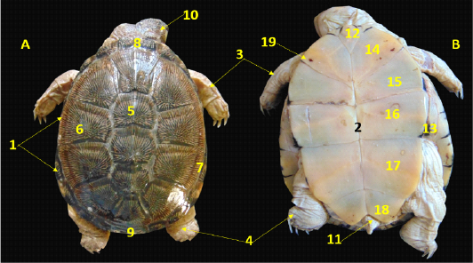
Figure 1: A. Dorsal view, B. Ventral view showing the external features of the female African helmeted turtle. 1. Carapase, 2.Plastron, 3.Forelimb, 4. Hind limb, 5.Vertebral scute, 6. Lateral costal scute, 7. Marginal scute, 8. Nuchal scute, 9. Supracaudal scute, 10.head, 11. Tail, 12. Gular Plastral scute, 13. Inguinal Plastral scute, 14. Humeral Plastral scute, 15. Pectoral Plastral scute, 16. Abdominal Plastral scute, 17. Femoral Plastral scute, 18. Anal Plastral scute, 19. Rathke’s pores
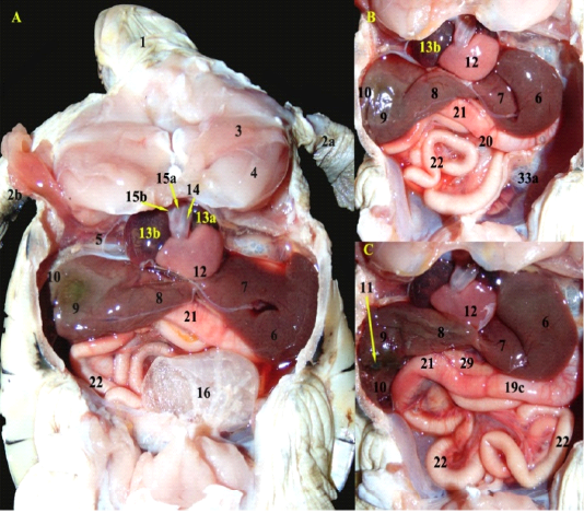
Figure 2: A; A photograph showing the ventral view of the coelomic cavity of African side neck turtle (fresh specimen) B; The urinary bladder was totally reflected, C; The right lobe of liver was slightly reflected.)
The Gastrointestinal Tract and Associated Organs
Pharyngeal openings: The soft palate was absent therefore the oral cavity and pharynx formed the oropharynx. The openings of the pharynx were six openings; paired internal choanal opening (fig.6A/3), paired opening of Eustachian tubes (fig.6A/4) on both sides of the roof of oropharynx caudally, single glottis (fig.6A/6) as a vertical slit and aditus esophagus. Also, rostral to the hard palate; dense rows of villiform papillae (fig.6A/2) in the form of inverted V-shape located in the upper jaw.
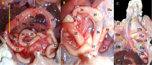
Figure 3: A; Photograph showing the pancreas parts after slight reflection of duodenum upward and rostrally, B; Magnified spleen in relation with the pancreas, C; The thoracic muscles were removed, neck was dissected and heart was removed.
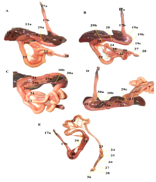
Figure 4: Photographs showing the Separated GIT; A; Ventral view, B; reflection of left lobe of liver, C; reflection of duodenum upward, D; parietal surface of stomach in relation to spleen, E; liver was removed.
Legend of figs 2, 3 &4: 1. Head, 2a. Left forelimb, 2b. Right forelimb, 3. Deltoideus muscle, 4. Supracoracidus muscle, 5. Horizontal septum, 6. Left lateral lobe of liver, 7.left medial lobe,8. Middle lobe, 9. Right medial lobe, 10. Right lateral lobe, 11. Gall bladder, 11a, bile duct, 12. single Ventricle, 13a. Left atrium, 13b. Right atrium, 14. pulmonary trunk, 15a. Left Aorta, 15b. Right aorta, 16. Urinary bladder, 17a. first third of Oesophagus, 17b. Second two thirds of esophagus,18. Gastro oesophageal sphincter, 19. Stomach, 19a. cardiac region, 19b. fundic region, 19c. pyloric region, 20. Pyloric sphincter, 21. Duodenum, 22. Jejunal loops, 23. Ileum, 24. Cecal bulb, 25.Transverse colon , 26. Descending Colon, 27. Rectum 28. Cloaca , 29. Pancreas, 29a.head of pancreas, 29b.body of pancreas, 29c. Tail of pancreas, 29d, pancreatic duct, 30. Spleen, 30a, base of spleen, 30b. apex of spleen, 31. Trachea, 32a. Left primary bronchus, 32b. Right primary bronchus, 33a. Left lung, 33b. Right lung, 34. Left kidney, 35. Left ovary. 36. Vent.
Gastrointestinal tract: The esophagus (fig.3C/17a&B) was a long Musculomembranous tube ended after the bifurcation of the trachea, it passed dorsally and slightly to the left the trachea, then to the left to reach the cardia. The stomach (fig.4D/19) was simple and fusiform, located totally on the left side. The stomach divided into; cardiac, fundic (fig.3C/19a&b) and pyloric region (fig.2C/19c). The pyloric sphincter (fig.2B/20) was short, smooth, and constricted to the left. The duodenum (fig.2/21) was short organ mostly at the right coelomic cavity, with a larger diameter than the pylorus (fig.5B) and ended at the level of the gall bladder. The jejunum (fig.3A/22) was a long-convoluted tube with constant diameter started at the right coelom to the left coelom; its diameter was lesser than that of ileum. Both jejunum and ileum internally had very small longitudinal folds. The ileum (fig.3A/23) was a short tube which opened into the cecum with the ileocecal valve (fig.5D/10). The small cecal bulb (fig.3A/24) was present at the right of the median plane. The large intestine composed of; a transverse colon (fig.3A/25) as a large pouch, from the right to the left of the median plane; dorsal to the small intestine, then it converted into the descending colon (fig.3B/26) as a short, wide and slightly to the right of the median plane. It followed by the narrower rectum (fig.3B/27), then finally into the cloaca. The cloaca (fig.6D/23) ended externally as the vent. The cloaca composed of coprodeum, urodeum and proctodeum.
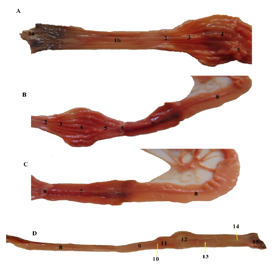
Figure 5: A, B, C &D: Photographs showing the opened gastrointestinal tract parts showing their mucosa.1a. 1st third of Oesophageal mucosa (pigmented and papillated with small pointed cones), 1b. Last two thirds of Oesophageal mucosa with numerous thin longitudinal folds, 2. Gastro oesophageal sphincter mucosa with small longitudinal folds without papillae. 3. Cardiac region of stomach with thin longitudinal mucosal folds, 4. Fundic part with thick corrugated longitudinal folds, 5. Pyloric region with few longitudinal folds. 6. Pyloric sphincter mucosa without any folds. 7. Duodenum smooth, dark red colored mucosa, 8. Jejunal mucosa, 9. Ileum mucosa, 10. Ileocecal valve mucosa, 11. Cecal smooth mucosa (saccular region), 12. Transverse colon smooth mucosa, 13. Descending colon mucosa with rectilinear folds, 14. Rectal smooth mucosa, 15. Cloacal mucosa.
GIT associated organs:
The liver
It was the largest abdominal organ, dark red in color and extended transversely in the center of the coelomic cavity. It was partially divided with moderated fissures into five lobes; divided right, left, and middle lobes (fig.2A/8). A small glistening rounded gall bladder (fig.2C/11) lodged between the right lateral (fig.2C/10) and medial lobes (fig.2C/9) at their visceral surfaces toward the caudal border. The left lobe related to the stomach, spleen and the right lobe related to the duodenum. The middle lobe was small heart shaped.
The compact pancreas
It (fig.2C/29) was pink to orange in color, began at the pylorus and passed parallel to the duodenum then ended just after the duodenum. It had three parts: narrow wedge-shaped head (fig.3A/29a), wide long body (fig.3A/29b), and small triangular tail (fig.3A/29c). A pancreatic duct (fig.3A/29d) arose from the last part of the pancreatic body.
The spleen
The spleen (fig.4E/30) was pear shaped, had faint red coloration, it had a large base (fig.3A/30a) related to the lesser curvature of the stomach, a smaller body and a pointed apex (fig.3A/29b).
The Respiratory System
Trachea (fig. 6B/7) had complete cartilaginous circular rings then bifurcated at the level of thoracic inlet and dorsal to the heart into the extra pulmonary bronchus (fig.6B/9) which passed for a distance outside the lung then changed to the pulmonary bronchus into the corresponding lung at its ventromedial surface. The Lungs (fig. 6C/10) were large sponge vascular sacs with many septae, which attached dorsally and extended caudally till reached to genitalia, so the posterior angles of the lungs tapered and lodged between the kidneys and ovaries.
The Urinary System
It composed a pair kidneys and ureters as well as three urinary bladders. Kidneys (fig. 6D/15, fig.7/1) were elliptical in shape, brownish in color, located dorsally so it was retro coelomic. Its dorsal surface was convex smooth, while it’s ventral surface was straight, lobulated and carried a segmental artery arose from the aorta which given it a special appearance, then covered by pleuroperitoneal membrane. Both kidneys located at the same level in the pelvis region in relation to adrenal gland, ovary, and lung. Adrenal glands (fig. 6C/16, fig.7/3) were paired small with orange coloration which adhered to the medial border of both kidneys. The two ureters (fig.6D/17) passed from both kidneys into the urodeum of the cloaca from its dorsaolateral aspects. A pear sac like urinary bladder (fig.2A/16) presented in the midline of the pelvis dorsal to the pubis and it opened directly in the ventral floor of the cloaca. In addition to other two accessory urinary bladders appeared laterally at the neck of the main urinary bladder.
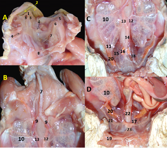
Figure 6: A. Roof and floor of oropharynx; B, C, D; anterior, middle, posterior parts of coelomic cavity of female African side neck turtle, respectively. 1. Hard palate, 2. Villiform papillae, 3. Internal Choanae, 4. Opening of Eustachian tube, 5. Tongue, 6. Glottis.7. Trachea, 8. Cut oesophagus, 9. Extra pulmonary bronchus, 10. Lung, 11. posterior angle of lung, 12. Left aorta 13. Right aorta, 14. Dorsal aorta 15. Kidney, 16. Adrenal gland, 17. Ureter, 18. Segmental artery, 19. Urinary bladder (reflected), 20. Ovary, 21. Oviduct, 22. Descending Colon, 23. Cloaca.
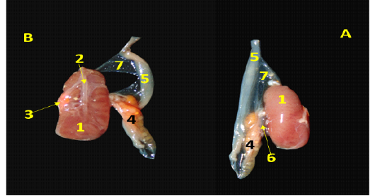
Figure 7: Photographs showing kidney in relation with ovary, A. Dorsal view; B. Ventral view, 1. Kidney, 2. Segmental artery, 3. Adrenal gland, 4. Ovary, 5. Oviduct, 6. Mesovarium, 7. Mesosalpinx.
The Juvenile Female Reproductive System
It consisted of a pair of ovaries, two oviducts, mesovarium and a mesosalpinx. The immature ovaries (fig.6C/20, fig7/4) on both sides were elliptical elongated granulated in appearance with yellow coloration and presented in contact with the kidneys, the mesovarium suspended the ovary to the coelomic cavity. The oviduct (fig.6D/21, fig.7/5) was narrow zigzag tube with thin wall, located dorsal to the urinary bladder and extended from the cranial pole of the ovary and suspended by mesosalpinx beyond the ovary till opened in the cloaca laterally.
Discussion
The carapace is a keratinized scute layer covering the underlying united bones and its nomenclatures are in a line with McArthur et al. (2004). while Wyneken (2001) described the shell is more leather like skin.
In our study we observed that the ventral coelomic area and dorsal retro-coelomic area divides by the horizontal septum. Due to the lack of diaphragm in the sea turtles and all reptiles, a single coelomic cavity included all their viscera (Baptistotte, 2014; Baldini, et al., 2019), which divides into a pericardial cavity and a common peritoneal cavity (Legler, 1993). Also, our work confirms a retro-coelomic area includes the lungs, kidneys and gonads (McArthur et al., 2004). In the current work the heart is three-chambered immediately above the plastron in the midline, cranial to the liver which in a line with (Wyneken 2001; McArthur et al. 2004; Baldini et al. 2019) while the former author added that two aortas and a pulmonary trunk get up from the anterior and ventral part of the heart.
We asserted the soft palate is absent in the pharynx which confirmed by McArthur et al. (2004). In accordance with (Wyneken, 2001) the paired choanae situates dorsal to the glottis, also the openings to the eustachian tubes locates at the caudal lateral parts of the palate. The current examination confirmed that the villiform papillae in the mouth is present which in accordance with McArthur et al. (2004).
Concerning the GIT, the esophagus is dorsal to the trachea then passes to the left to reach the stomach, in accordance Magalhães et al. (2012). Conversely Wyneken (2001) detected that the esophagus passes to the right of the trachea, in green turtles, there is a crop, also, in cheloniids, and the esophagus is S-shaped. In our work, the esophagus has thin and conical papillae at first part only, per- contra, Magalhães et al. (2012) stated that the last part of the esophageal mucosa increases in papillae. In agreement with the description of Magalhães et al. (2012), Gastro esophageal sphincter is devoid of papillae, while in some species a pouch like esophageal diverticulum is present. The stomach is simple and fusiform, locates on the left side, simulating the observations of McArthur et al. (2004). While Houck et al. (2019) detected that the stomach is tubular. The pyloric sphincter is away from the level of the midline of the liver. However, Houck et al. (2019) approved that the pyloric sphincter is near midline. In the present study, the pyloric sphincter is short, smooth. Per- contra Magalhães et al. (2012) revealed that it has longitudinal folds.
The duodenum is a short organ situated mostly at the right coelomic cavity these findings simulated Magalhães et al. (2012). While Houck et al. (2019) explained that it has cranial flexure. The duodenum has a plain surface internally, simulating the findings of Wyneken (2001), while in some species, it is honeycomb in shape, and reticular in others (Magalhães et al., 2012). We investigated that the diameter of the duodenum differs from that of the pylorus and the jejunum. Conversely, Magalhães et al. (2012) cited that the small intestine has a constant diameter. In accordance with Baldini et al. (2019) as they detected that the jejunum observes bilaterally. Similar to the observations of Houck et al. (2019).
The jejunum and ileum internally has very small longitudinal folds, also the iliocecal sphincter is smooth internally, while Magalhães et al. (2012) revealed that in the jejunum and ileum h honey-comb reticular folds, in addition to the ileocecal sphincter of D. coriacea a small lateral pouch.
In the line with the observations of Magalhães et al. (2012) the descending colon has rectilinear folds. The present study as well as those of Houck et al. (2019) a small cecal bulb locates ventrally in the right coelom, in addition to both transverse and descending colon. Our results confirmed that the rectum joins the cloaca then externally as a vent, where the cloaca composes of coprodeum, urodeum and proctodeum which in accordance with Wyneken (2001).
The present study as well as those of Gbadebo et al. (2014), in the African side neck turtle, a middle lobe of liver is present. While Moura et al. (2012) revealed that this lobe was named central lobe. Also, Wyneken (2001) named it connecting strips. Per- contra Machado Júnior et al. (2005) investigated that liver divides into; right lateral, median, square, left lateral and caudate with a papillae process. Moreover, Moura et al. (2017) generated that it composes of four lobes; divided right and left lobes. The present study convinced that the left lobe is greater than the right one, contradictory with Wyneken (2001). In accordance with Wyneken (2001) the liver is dark brown and the largest visceral organ. Per- contra, Moura et al. (2017) conducted that it is light brown organ with black spots.
In our study we find that the pancreas has three parts: head, body, and tail. A pancreatic duct arises from the last part of the pancreatic body and opens just before the end of the duodenum and. Conversely, Wyneken (2001) mentioned that its shape is an irregular strip and the pancreatic duct is difficult to locate. Moreover, McArthur et al. (2004) surveyed that a larger number of pancreatic ducts present. Similar to the observations of Wyneken (2001) that the pancreas is pink to orange in color, While McArthur et al. (2004) generated that it is pale. In agreement with Stahl (2003) in the snake-headed turtle as their pancreas correlates with the spleen proximal part. While, in the South American side-necked river turtle there is no interrelation between the pancreas and the spleen. Also, Wyneken (2001) stated that spleen relates to distal end of pancreas. In our study, the spleen is pear shaped, while Baldini et al. (2019) revealed that the spleen is oval.
Concerning the respiratory system, the trachea divides into paired bronchi, simulating Mans et al. (2013). Lungs are sponge sac as mentioned by (McArthur et al., 2004). We disagreed with McArthur et al. (2004) as our results mentioned that the posterior angles of the lungs are tapering and enters between the kidneys and ovaries but does not cover the kidney. And in a line with Silva et al. (2010) as they noted that the kidney is present in close contact with the oviduct in females.
In the urinary system, Silva et al. (2010) revealed that a pair of asymmetrical kidneys are found which was unlike our result that asserted both kidneys are symmetric as mentioned in lizard (Yokota et al., 2005). In our study, the turtle kidneys are elliptical while it was elongated in lizards and crocodiles, but narrow in snakes (Silva et al., 2010). In accordance with Yokota et al. (2005) the kidney of turtle and domestic fowl surrounds by segmental artery. The two ureters penetrate the urodeum dorsolaterally, similar to the observations of (McArthur et al. 2004), as the ureters open in the urodeum at approximately 10 and 2 o’clock positions in our study, at transverse cross section. In the current study there are one main urinary bladder and two accessory bladders, but Crocodilians and snakes lack the presence of urinary bladders (Wyneken, 2013). The urinary bladder is a pear sac in shape with highly thin elastic membrane as (Baldini et al., 2019). The urinary bladder opens directly in the floor of cloaca so the contents of the cloaca may be redirect to the urinary bladder according to (Wyneken, 2013).
the continent of the female genital system are two ovaries, two oviducts as well as mesovarium that in accordance with (Sánchez-Ospina et al. 2014, Spadola et al. 2017), The latter author denie the presence of mesosalpinx, While Takami (2017) confirms the presence of it. Unlike our description Sánchez-Ospina et al. (2014) noted that ovary has a transparent to white color and the mesovarium has black pigmentation, conversely, we found that the ovarian color is yellow and the mesovarium is transparent. We confirmed our results as Wyneken (2001) that the ovary locates medial to the oviduct which passes anteriorly then curved caudally. Also, Spadola et al. (2017) added that the immature ovary has not prominent ovarian follicles as we noted in this study.
Conclusions
Unlike mammals, the obvious conclusion is the absence the diaphragm leads to nonattendance of the pleural and abdominal cavities which substitute by the presence of ventral coelomic area and dorsal retro-coelomic area divides by the horizontal septum (transverse pleuroperitoneal membrane). Concerning the digestive tract and associated organs; the openings of the pharynx are six openings which differ from the fowl. The diameters and length of the small intestine parts differs. The liver has five lobes; also a gall bladder is present. The compact pancreas has head, long body and triangular tail. The lungs are very long covering more than half of the carapace’s length till reaches caudally to ovary and the kidneys. Both kidneys are elliptical locate at retro coelomic area at the same level in the pelvis region. In the juvenile female; the paired immature Ovaries are elliptical elongated granulated in appearance with yellow coloration.
Data availability
Data sharing is not applicable to this article as no new data were created in this study.
Authors contribution
Both authors have made substantial contributions to the research design, or the acquisition, and to drafting the manuscript or revising it; and that all authors have reviewed and approved the last submitted version.
Funding
The authors ́ research included into this article did not receive any specific grant from funding agencies in the public, commercial, or not-for-profit sectors.
Conflict of interest
No conflict of interest
References


