Advances in Animal and Veterinary Sciences
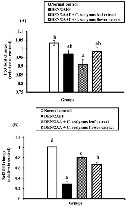
Effect of C. scolymus leaf and flower hydroethanolic extracts on kidney (A) p53 and (B) Bcl-2 mRNA gene expressions.
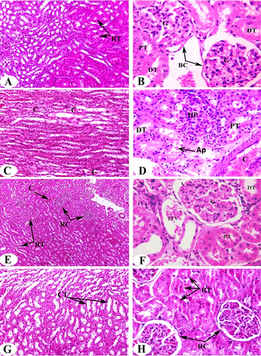
Photomicrographs of H and E stained kidney sections of rats in the experimental groups. Photomicrographs (A, B) clearly shows typical glomerulous histological structure (G), proximal (PT) and distal tubules (DT) in normal rat. Photomicrographs of kidney sections of DEN/2AAF show congestion (C) in the intertubular blood vessels of renal medulla (Photomicrograph C), hyperplastic proliferation of the glomerulus (HP), apoptotic cells (Ap) and congested renal artery (C) in the cortical region (Photomicrograph D). Photomicrograph of kidney section of DEN/2AAF-administered rat treated with C. scolymus leaf extract showing nearly normal renal tubules (RT) (Photomicrograph E), glomerulus (G), proximal (PT), distal tubule (DT) and inter-tubular vein (ITV) (Photomicrograph F). Photomicrograph of kidney section of DEN/2AAF-administered rat treated with C. scolymus flower extract showing nearly normal collecting tubules (CT) (Photomicrograph G) (X 200), renal tubules (RT) and corpuscles (RC) (Photomicrograph H) (X 400).


