Advances in Animal and Veterinary Sciences
Research Article
Adv. Anim. Vet. Sci. 8(s3): 47-55
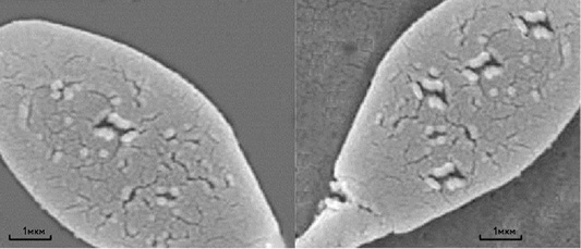
Figure 1
Scanning electron microscopy of thawed sperm cells of a stallion without the addition of mycotoxin (left) and with the addition of mycotoxin (right). Scale segments - 1 µm.
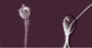
Figure 2
Light microscopy of thawed bull sperm cells with the addition of Zearalenone and the T-2 toxin (sperm cells with damaged acrosomes are shown). Magnification 100×.


