Advances in Animal and Veterinary Sciences
Research Article

Studying the Factors Affecting the State of Cattle Hoof Horn
Tatiana Babintseva*, Ekaterina Mikheeva, Alexander Shishkin, Vitaly Yudin, Ekaterina Klimova, Konstantin Shklyaev
Izhevsk State Agricultural Academy, Studencheskaya Street, 11, Izhevsk, 426069, Russia.
Abstract | In this study, we evaluated the incidence of necrobacteriosis in cattle based on the environmental factors and climatic features in the Udmurt Republic. the distal extremities pathology structure in cattle was determined in several farms of the Udmurt Republic, where an average of 25-27.5% of the animals had musculoskeletal lesions. The dynamics of the necrobacillosis epizootic process in cattle were studied from 2011 to 2016 and the highest incidence rates were recorded in 2015 with 8.86%. Our results revealed that the pathology of the gastrointestinal tract, in particular, disruption of the microbiocenosis of the rumen and subacute and chronic enter colitis, in combination with unbalanced feeding, affected the degree of damage to the hoof horn. Likewise, the level of metabolic disorders in animals with lesions of the hoof horn was determined. The relationship between the environmental factors, climatic characteristics in the Udmurt Republic, pathology of the gastrointestinal tract, unbalanced feeding and the degree of lesion of the hoof horn has been established.
Keywords | Cattle, Diseases of the distal extremities, Deformation of the hoof horn, Metabolic disorders, necrobacteriosis
Received | August 01, 2020; Accepted | August 7, 2020; Published | August 28, 2020
*Correspondence | Tatiana Babintseva, Izhevsk State Agricultural Academy, Studencheskaya Street, 11, Izhevsk, 426069, Russia; Email: ariadna-357@mail.ru
Citation | Babintseva T, Mikheeva E, Shishkin A, Yudin V, Klimova E, Shklyaev K (2020). Studying the factors affecting the state of cattle hoof horn. Adv. Anim. Vet. Sci. 8(s3): 11-17.
DOI | http://dx.doi.org/10.17582/journal.aavs/2020/8.s3.11.17
ISSN (Online) | 2307-8316; ISSN (Print) | 2309-3331
Copyright © 2020 Babintseva et al. This is an open access article distributed under the Creative Commons Attribution License, which permits unrestricted use, distribution, and reproduction in any medium, provided the original work is properly cited.
INTRODUCTION
Previous studies revealed that diseases of the distal extremities, including necrobacteriosis, are one of the leading pathologies of cattle in terms of the prevalence and economic damage to agricultural enterprises as a result of. intensification of animal husbandry (Ivanov et al., 2013; Lopatin et al., 2014; Samolovov et al., 2011; Khuzin, 2015; Dippel et al., 2009).
The nature of diseases of the distal extremities is polyetiological (Manske and Hultgren, 2002; Cruz et al., 2001; Biowey, 2005). The main reasons for the development of these diseases include violations of the veterinary and sanitary rules for keeping animals, increased humidity in the premises, untimely removal of manure, unbalanced vitamin, micro- and macroelment intake, fiber deficiency, selection of highly productive animals. Also, this can be caused by the herd rearrangement and the introduction of new, especially imported, livestock into the herd beside climatic factors that might play a significant role. For example, high humidity, as well as dry and hot summers affect the condition of the skin and hoof horn. (Ivanov et al., 2013; Melnikova, 2013; Miheeva et al., 2013) A high percentage of culling due to damage to the distal extremities was recorded on farms and the proportion of animals with such lesions can reach up to 60% out of the total number of culled animals (Ivanov et al., 2013; Melnikova, 2013; Miheeva et al., 2013; Khuzin, 2015; Smith and Thornton, 1993). Housing large numbers of animals in limited areas, keeping livestock on a leash, absence of active exercise, and especially with slotted cold concrete floors without bedding leads to the loss of strength in hoof horn, becomes friable, creases and deformation of the horn will occur consequently (Lopatin, and Samolovov, 2014; Tikhonova et al., 2012). The tissues of the coronary band and the interhoof fissure undergo maceration and, therefore, become susceptible to opportunistic microflora and the occurrence of necrobacteriosis (Lopatin, 2014; Tikhonova et al., 2012).
The highly concentrated type of feeding has a significant effect on the spread of hoof diseases in highly productive cattle (Ivanov et al., 2013; Melnikova, 2013; Miheeva et al., 2013). The use of feed containing a significant amount of acetic and propionic acids and crude protein with a deficiency of fiber in the diet leads to a sharp decrease in the number of cellulolytic microflora in cows and an increase in the number of microorganisms with proteolytic properties (Smith and Thornton, 1993; Mulling et al., 1999). A long-term deficiency of micro and macroelements, especially calcium, zinc, sulfur, and copper, contributes to the development of osteodystrophy, pathology of the skin and its derivatives, blood vessels and alveoli, dysregulation of intracellular processes, disruption of the permeability of cell membranes, and a decrease in the functional activity of immune cells. A large amount of histamine enters the body of cows, which, after being released from the grains, form labile bonds with the proteins of the hoof horn, which contributes to the development of rheumatic inflammation of the hooves, leading to massive deformities. Inflammatory processes in the hooves area are often accompanied by compression of the skin matrix and impaired blood supply, which leads to tissue necrosis (Samolovov and Lopatin, 2011; Tolkachev, 2015; Vasiliev et al., 2016). Several studies correlate the occurrence of hoof pathology with metabolic disturbances and, accordingly, decrease in general resistance and tissue resistance in the setting of the development of secondary immunodeficiency states (Samolovov and Lopatin, 2011; Lopatin and Samolovov, 2014, Mikheeva et al., 2013; Burdov et al., 2015). The aim of the study is to determine the causes of diseases of the distal extremities, including necrobacteriosis, in the Udmurt Republic (Western Cis-Ural region). The following tasks were accomplished during this research: study the cattle necrobacillosis incidence rates in the Udmurt Republic, determination of the nature of the lesions of the distal extremities and study the degree of impact of the climatic features, maintenance and feeding conditions on the occurrence of hoof diseases.
MATERIALS AND METHODS
The study was carried out after approval from Department of Infectious Diseases and Pathological Anatomy of the inter-faculty educational and scientific laboratory of the Federal State Budgetary Educational Institution of Higher Education “Izhevsk State Agricultural Academy” and the farms of the “Rossiya” LLC and “Vera” LLC in the Udmurt Republic in 2012-2016. This study was included1,363 cattle’s. Blood, organs, and tissues, ruminal contents, feces samples, reporting documentation of the Main Directorate of Veterinary Medicine of the Udmurt Republic, the regional animal disease fighting station, the Udmurt Center for Hydrometeorology and Environmental Monitoring, and the annual reports of the “Rossiya” LLC and “Vera” LLC (Udmurt Republic) were used as the research material.
Clinical and orthopedic study of dairy cattle was carried out according to the following scheme: (1) examination at rest (the positioning and setup of the limbs, the nature of the setup and the state of the hooves were recorded); (2) examination of movement (the type, degree, and nature of lameness) were recorded and (3) palpation of the distal extremities (tissue elasticity and sensitivity, size of the lesion and its nature) were determined. According to the results of examination, cows were divided into clinically healthy animals and animals with lesions of the distal extremities.
The contents of the rumen were taken using a probe two hours after feeding to the study of ruminal digestion. Using universal test strips, pH was measured, the amount of ruminal microflora was studied using the Goryaev chamber, mobility was measured in a hanging drop, and activity was measured in a sample stained using methylene blue. The digestibility of the feed was determined by the composition of feces. Feces were taken from the rectum and washed using a sieve to determine the size of undigested feed particles. A morphological study of the gastrointestinal tract of dairy cows was carried out and tissues of the rumen and small and large intestines were used for histological studies. To determine the quality of the hoof horn, material was taken from animals with a mild, moderate, and severe degree of damage of the hooves. Total protein content in blood serum was measured using the refractometric method (Kondrakhin et al., 1985), and the content of protein fractions in blood serum was studied using by the nephelometric method (Kondrakhin et al., 1985). Biochemical parameters of blood serum (content of glucose, alkaline phosphatase, calcium, phosphorus, zinc, iron) were determined using the Flexor E automated biochemical analyzer (Netherlands) and Stat Fax 1904 plus (Vital Diagnostix test systems, Russia).
RESULTS AND DISCUSSION
A total of 643 animals were examined during the surgical medical examination of cattle in the “Vera” LLC milking herd that revealed that25% of the cows had extremities lesions: 53% had hyperextension of the tendon-ligamentous apparatus; 41% had deformed hoof horn; 50% had acute and chronic inflammation in the hooves area. On the other hand, a total of 720 animals were examined during the surgical medical examination of cattle at the “Rossiya” LLC where 27.2% of animals had lesions of the distal extremities: 45.4% had hyperextension of the tendon-ligamentous apparatus; 50.5% had deformed hoof horn; 54.6% had acute and chronic inflammation in the hooves area.
Our observations revealed that mostly the hind limbs were affected, that can be explained by an increased load on the back of the body and prolonged contact with the urine and feces. Moreover, the Udmurt Republic has a permanent distressed status for cattle necrobacillosis. During 2011, according to General Directorate of Veterinary Medicine of the Udmurt Republic, the incidence rate for cattle necrobacillosis was 1.95%, and there were three distressed locations at this time as shown in Figure 1.
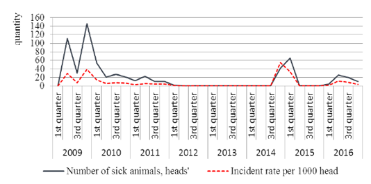
Figure 1: Data on the distressed status for necrobacteriosis in cattle in the Udmurt Republic in 2011-2016.
After 2011, in consequence of measures to eliminate necrobacteriosis, all indicators decreased and reached zero while in 2014, animals with necrobacteriosis began to be reported. By 2015, the incidence reached 8.86%, generally, the peak of sick animals was reporting in the 2nd and 4th quarters of 2011 and 2014 and in the 1st quarter of 2012 and 2015; the middle of autumn, winter, and the beginning of spring.
The Udmurt Republic is located in the middle northern latitudes of the western part of the Middle Urals and is characterized by a temperate continental climate with cold snowy winters and relatively warm summers. The average annual temperature ranges from 1.0 to 2.5ºC; July is the warmest month of the year (17.5-19ºC), and January is the coldest month (-14-15ºC). The total amount of precipitation during the year is 638 mm; 323 mm of precipitation falls during the warm season (May-September), the largest amount of which falls in June (78 mm). Natural and climatic conditions might have a significant impact on the development of diseases for the musculoskeletal system in cattle. For example, the increase in the number of animals newly infected with necrobacteriosis in 2011 may be associated with high humidity during this period: 108.1 mm of precipitation fell in June when the normal amount is 78 mm (Figure 2).
In July of 2011, 147 mm of rain fell, which was 207% more than normal, and that contributed to spoilage of feed and a decrease in its nutritional value during its procurement, which could lead to the increase in the percentage of culling of animals due to diseases of the distal extremities in 2012. Likewise, high humidity might contribute to the higher the incidence of necrobacteriosis in the 4th quarter of 2014. In October, 114.3 mm of precipitation fell (climatic normal is 62 mm). The average monthly temperature was 0ºC, which was 2.7ºC lower than the climatic normal (Figure 3).
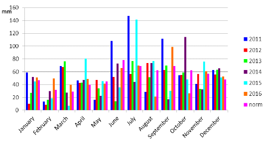
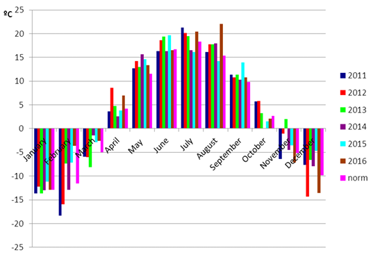
Moreover, during 2016, the climatic conditions were favorable for the spread of necrobacteriosis infection, the average temperature in February was -3.6ºC, which was 8ºC higher than the climatic normal (-11.6ºC), with 49.7 mm of precipitation – 155% more than the normal (32 mm). In March, the temperature was -2.6-2.4ºC higher than the climatic normal (-5ºC), 39.8 mm of precipitation fell (the norm is 29 mm). In April, there was 48.5 mm of precipitation – 121% more than the normal (40 mm), and in September this indicator was 98.9 mm, which was 143% more than the normal.
In addition, in July of 2015, 141 mm of precipitation fell, which was 205% higher than the climatic normal, and the temperature was 16ºC, which was 2.2ºC lower than the climatic normal. This contributed to the procurement of feed of poor quality, which could be the reason for the necrobacteriosis outbreaks in 2016.
Also, violation of the rules of veterinary-sanitary control of livestock buildings was also one of the reasons for the development of diseases of the distal extremities, including necrobacteriosis. Increased humidity (condensate on the walls, beams, and pipes), untimely removal of manure, lack of litter, and crowded keeping of animals were found. The exercising area was in an unsatisfactory condition (the area was flooded in rainy weather, the dirt reached up to the level of the cows’ fetlock joint, sporadic nails and wire were found).
One of the main factors that contributed to the development of pathology of the distal extremities in cows was the unbalanced feeding of animals. Our study revealed that with the analysis of the level of feeding showed that the proportion of silage in the diet of dairy herd cows was up to 71.5% and the proportion of concentrates was 14.2%. Coarse feed accounted for only 8.7% as shown in Table 1, while the main used feeds were corn silage, grass-legume silage, rapeseed silage, and grass-legume grain-haylage. Samples of these feeds were analyzed by an accredited independent testing laboratory of the Udmurt sky Agrochemical Center JSC showed that the amount of metabolic energy in the feed was normal, while there was a 5-13% excess of protein, 7.8-27.7% excess of calcium, and an average of 12.5% excess of phosphorus. The ratio of calcium and phosphorus was 2.9:1 in hay, 2.62:1 in haylage, and 0.37:1 in silage.
Table 1: The diet of dairy cows in the “Rossiya” LLC.
| Feeds | Amount, kg | Proportion in the diet, % |
| Silage | 28-30 | 71.5 |
| Concentrates | 5.8 | 14.2 |
| Hay | 1.5 | 3.7 |
| Mill cake | 1.0 | 2.5 |
| Helm | 2.0 | 5.0 |
| Molasses | 1.0 | 2.5 |
| Salt | 0.2 | 0.9 |
An excess of calcium and phosphorus might lead to a shift in the ratio of these nutrients; in addition, an excess of calcium entails problems with the absorption of critical elements, such as zinc, manganese, phosphorus, and other minerals (Vasiliev et al., 2016). Our results demonstrated a significant deficiency of some feed nutrients: fiber deficiency averaged 19%, sugar deficiency was 6.6%, deficiency of crude fat was up to 50% on average. Sugar and crude fat deficiency can lead to a disruption in carbohydrate-fat metabolism, which in turn leads to acidosis, accumulation of ketone bodies, a decrease in the alkaline reserve of blood, as well as reduction of the animals’ productivity and, in particular, reduction of the mass fraction of fat in milk (Vasiliev et al., 2016). Periodically, in response to increase the acidity of feed mixtures, the presence of butyric acid in the silage, and mold formation will be recorded.
Rumen plays the main role in the digestion of feed. Our study assessed the condition of the rumen by its content in cows without signs of hoof damage (healthy) and animals with damaged distal extremities (diseased). In animals with hoof lesions, the rumen pH more often shifted to acidity (about 5-5.5), and less often to alcalinity (more than 7.0). A decrease in the number of active ciliates to 30-50% indicated a disruption of the microbiocenosis. In clinically healthy cows, the pH of the rumen was in the range of 6.2-6.7. However, the percentage of active ciliates was in the range of 50%, which may be associated with the presence of concentrates and a high fineness of the feed.
The feces of the cows with visible signs of hoof damage had a dark green color, a creamy consistency, 50% of the samples foamed, which explained by increasing in the enzymatic activity of microorganisms of the large intestine (Vasiliev et al., 2016). Meanwhile, the undigested feed residue contained whole grains of herbs (cereals, corn), seeds of meadow plants, and long fiber particles 2-5 cm in size, which indicated a disruption of the rumen microbiocenosis was noticed in cows with visible signs of hoof damage compared to healthy animals, undigested components in the intestinal contents included plant particles less than 0.5 cm in size, corn shells, and single cereal grains.
During the histological examination of the rumen wall, moderate blood filling of the vessels of the mucous layer was observed and slight infiltration by lymphocytic elements was determined in healthy animals and cows with a mild degree of hoof damage. The mucosal epithelium was desquamated in places. Slight infiltration by the cells of the lymphoid series was observed in the basal part of the mucous membrane of the small intestine. The epithelium of the apical part of the villi was focally desquamated (Figure 4a). Additionally, slight diffuse eosinophilic cell infiltration was determined in the large intestine. In several animals, moderate hyperemia of the vessels of the mucous layer was observed (Figure 4b).

Figure 4: The mucous membranes of small (A) and large (B) intestines in cows with mild hoof lesions. Staining with hematoxylin and eosin. 100 x magnification.
In cows with a moderate degree of the distal extremities damage, microscopic examination of the rumen wall revealed hyperemia of the blood vessels in the mucous and submucosal layers, their endothelium proliferated, and lymphocytic infiltrates were observed in the periarterial zone. The epithelium of the mucous membrane was desquamated in places; the papillae were deformed which in accordance with previous finding Babintseva (2018).
Histological examination of the small intestine in cows with a moderate degree of hoof damage revealed hyperemia of the blood vessels in the submucosal layer, their endothelium proliferated, and an accumulation of lymphocytes and eosinophilic granulocytes was found in the adjacent area. The villi were unevenly thickened, a part of them had club-shaped thickenings, a part of them merged with each other, blood filling of the capillaries was moderate. The stroma of the villi was infiltrated by eosinophilic granulocytes and lymphocytes. The intestinal epithelium underwent metaplasia and had a large number of goblet cells. The marginal epithelium of the villi was partially desquamated. Lymphoid nodules were detected in the mucous layer (Figure 5).
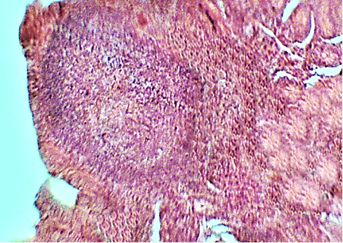
Figure 5: The mucous membrane of the small intestine in cows with moderate hoof lesions. Staining with hematoxylin and eosin. 100 x magnification.
Microscopic examination of the walls of the small intestine in animals with severe lesions of the hooves revealed hyperemia of the vessels of the mucous and submucosal layers and infiltration by lymphocytic elements and eosinophilic granulocytes mainly in the mucous layer (Figure 6). Some villi were unevenly thickened, and some were shortened, deformed, and fused together. The marginal epithelium of the villi was partially desquamated. The cells of the intestinal glands appeared cystically dilated.
During the histological examination of the walls of the large intestine, we observed polymorphic cell infiltration of the mucous layer consisting of a large number of eosinophilic granulocytes and lymphocytes. Goblet cells appeared enlarged, intestinal glands showed signs of metaplasia. The villi were deformed, merged in places. Vessels in the submucosal layer had moderate blood filling, lymphoid infiltration was found around them.
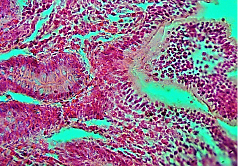
Figure 6: The mucous membrane of the small intestine in cows with severe hoof lesions. Staining with hematoxylin and eosin. 400 x magnification.
Therefore, a violation of the diet had a negative effect on the pH of the ruminal content and changed the composition of the microbiocenosis and the activity of the ciliates, which led to general acidosis and poor assimilation of nutrients of the feed. This affected the condition of the hoof horn. The lack of roughage in the diet led to an accelerated passage of feed through the rumen, as evidenced by poor digestibility of the feed. Foamy contents of feces indicated an increase in fermentation processes in the intestine. In animals with moderate and severe damage to distal extremities, we observed signs of subacute and chronic superficial and diffuse atrophic enteritis, as well as diffuse chronic colitis with signs of metaplasia of the intestinal epithelium and desquamation of the marginal epithelium of the villi (Table 2). Intestinal lesions exacerbated the poor assimilation of nutrients and micro and macroelements of the feed, leading to impaired synthesis of hoof horn. This was confirmed by biochemical studies of blood serum.
Table 2: Changes in the gastrointestinal tract in cows with varying degrees of lesion in the distal extremities.
| Lesions | Mild lesion | Moderate lesion | Severe lesion |
| Small intestine | insignificant infiltration with lymphoid cells | subacute superficial and focal atrophic enteritis | subacute and chronic superficial and diffuse atrophic enteritis |
| Large intestine | insignificant diffuse eosinophilic cell infiltration |
diffuse chronic colitis |
diffuse chronic colitis |
Analysis of the biochemical data from the blood samples from the “Rossiya” LLC obtained in the Mozhginsky interdistrict veterinary laboratory, revealed hypocarotinemia in 85.2% of animals; hypoglycemia in 97.1% of animals; low content of calcium in 56.9% of animals; low content of phosphorus in 18.4% of animals (above or below the norm); low content of protein in 23% of animals. On the other hand, the results of biochemical studies of blood samples from the “Vera” LLC were the following: Hypocarotinemia in 46.7% of animals; hypoglycemia in 99.4% of animals; low content of calcium in 13.3% of animals; low content of phosphorus in 40% of animals (above or below the norm); low content of protein in 26.7% of animals. In animals with hoof tissue damage, we detected hypocalcemia and hypophosphatemia where the level of Ca and P was 1.7 ± 0.29 mmol/L and 1.28 ± 0.5 mmol/L, respectively which was 1.5 times and 1.3 times lower than in healthy animals, respectively as shown in Table 3. The zinc content in the blood of cows with lesions of the distal extremities was 2.4 times lower than that of healthy animals and amounted to 8.35 ± 3.25 μmol/L Table 3.
Table 3: Biochemical blood parameters in cows with lesions of the distal extremities and clinically healthy animals.
| Parameters | Healthy | Diseased |
| Total protein content, g% |
7.87+2.08 |
7.4+1.86 |
| Ca, mmol/L |
2.6+1.86 |
1.7+0.29* |
|
Р, mmol/L |
1.63+0.42 |
1.28+0.5* |
|
Ca:Р ratio |
1.59:1 | 1.32:1 |
| Zn, μmol/L |
20.3+1.88 |
8.35+3.25*** |
| Fe, μmol/L |
4.5+0.63 |
4.3+0.33 |
*p < 0.05; **p < 0.01; *** p < 0.001 compared to the values in healthy animals.
Therefore, the increased number of diseased animals in the Udmurt Republic can be explained by high humidity caused by heavy rainfall and non-compliance with veterinary-sanitary rules for animal management that means exposure to adverse environmental factors led to the occurrence of lesions of varying severity in distal extremities.
The peak incidence of necrobacteriosis in 2015 coincided with the peak increase in rainfall and the preceding excessively wet period of fodder harvesting. Meanwhile, significant changes in the mineral composition of the blood were recorded in animals with tissue damage in the distal extremities, namely, pronounced hypophosphatemia, hypocalcemia, and hypozincemia. Likewise, unbalanced diet led to the disruption of ruminal digestion and development of subacute and chronic enterocolitis, which contributed to the disruption of the absorption of feed nutrients and the formation of the poor condition of hooves.
CONCLUSION
Our results revealed that the maximum incidence rates of necrobacteriosis in cattle in the Udmurt Republic were recorded in 2015 (8.86%). On average, 25-27.5% of animals had a musculoskeletal system lesion while hyperextension of the tendon-ligamentous apparatus, deformation of the hoof horn, and chronic inflammatory conditions were predominated. Meanwhile, there was a direct correlation between the incidence of necrobacteriosis and the climatic conditions at the Udmurt Republic. Finally, the use of feeds of poor quality with low nutritional value with a predominance of silage and concentrates led to impaired ruminal digestion and subacute or chronic enterocolitis, which might contribute to metabolic disorders and development of lesions of varying severity in the distal extremities of cattle.
Authors Contribution
All authors contributed equally to the manuscript.
Conflict of interest
The authors have declared no conflict of interest.
REFERENCES


