Advances in Animal and Veterinary Sciences
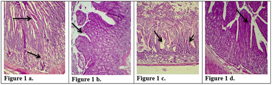
Description of histological changes in duodenum sections. a): Group III, negative control, animal feeding with saline solution. Normal appearance of microvilli, Brunner’s glands in duodenum. (200 X),(H & E)). b): Group II, positive control, animal feeding with E.histolytica cyst, hyperplasia and increases in microvilli and mucosal glands of duodenum. (200 X),(H & E). c): Group I a, section in the animal duodenum immunized by K-antigen, showing hyperplasia increases in length of intestinal microvilli (200 X),(H & E) d): Group I b, section in the duodenum shows the presence of hypoplasia of the intestinal microvilli with an increase in the mucous glands after infected immunized animal with E.histolytica (200 X)(H & E).

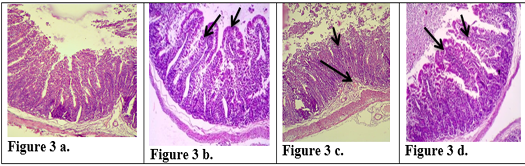
Description of histological changes in the jejunum sections. a): Group III, negative control, (200 X) (H & E). b): Group II, positive control, hyperplasia, (200 X) (H & E). c): Group I a, the section in jejunum shows increases in goblet cells and increases in length of mucosa region lining jejunum after an immunized animal with K-antigen (200 X) (H & E). d): Group I b, section in jejunum shows hyperplasia in the lymphatic glands (peyer’s patch) L.N. of submucosa with germinal center this refers to induce immunity (100 X) (H & E). e): Group I b, section in jejunum after infection with E.histolytica shows simple degenerations in the cells surrounding microvilli with filtration of inflammatory cells inside the cavity of intestinal microvilli and decrease in goblet cells (200 X)(H & E).
Description of histological changes in ileum sections. a): Group III, negative control animal feeding with saline solution, normal appearance of ileum (200 X)(H & E). b): Group II, positive control, animal feeding with E.histolytica cyst, (200 X)(H & E). c): Group I a, after injection with k-antigen the section in the ileum shows increases in length of intestinal microvilli and apoptosis in epithelial cell lining microvilli (200 X)(H & E). d): Group I b, section in jejunum after infection with E.histolytica shows simple degenerations in the cells surrounding microvilli with filtration of inflammatory cells inside the cavity of intestinal microvilli and decrease in goblet cells (200 X) (H & E)
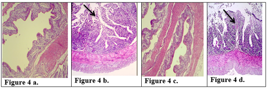
Description of histological changes in colon sections. a): Group III, negative control, normal appearance of colon (200 X) (H & E). b): Group II, positive control.(200 X) (H & E) submucosal ulceration. c): Group Ia ,section in the colon looks like normal and there is no hyperplasia in lining of colon mucosa after immunized animal with K-antigen (200 X) (H & E) d): Group I b, colon section shows superficial mucosal ulceration with heavy filtration of inflammation cells in and between folded intestinal microvilli after infected immunized animal with E.histolytica (200 X) (H & E).


