Advances in Animal and Veterinary Sciences
Case Report

Post-Mortem Examination of a Female Elephant Suspected of Having Degenerative Joint Disease: A Case Report
H. M. Usman Saddiq1*, Rana Haider Ali1, Muhammad Tahir Amjad2, Shahid Jaleel2, Syed Manuchahar Ali2, Noor Fatima1, Sami Ullah1
1University of Veterinary and Animal Sciences Lahore, Pakistan; 2Riyadh Zoological Garden, Al Malaz Ar Riyadh, Saudi Arabia.
Abstract | The musculoskeletal impairments are one of the major health challenging issues of captive elephants in zoos. This is a case study of 45 years old female African Elephant (Loxodonta africana) named Laila, kept at Riyadh Zoological Garden, Al Malaz Ar Riyadh, Saudi Arabia. There were total 3 female African elephants in the zoo and Laila was the eldest one. The elephant Laila was fed on fresh berseem and leaves, lucern hay, rhodes grass, pellets, molasses, rice, seasonal vegetables and fruits (Banana, Apple, and Orange). Facility of adequate water was provided by keeping drums of 100 litres capacity. The elephant was captivated at night quarters having cemented floor while during day time it was allowed to walk on soil. Usually one hind limb of the elephant was tethered with chain. The animal was found falling down repeatedly and unable to bear weight on limbs while attempting to stand up. Walking Activity was gradually decreased and ultimately total reluctance to walk. Bones were malformed grossly and splaying was evident. The elephant was suspected for gradual aging and related Degenerative Joint Disease. The animal was on continuous supportive medicines from the last few months. The animal was also under hot water hydrotherapy for quicker recovery from Degenerative Joint Disease. At the end, the elephant died when constant pain and lameness became unresponsive to protracted treatment. Post-mortem examination of elephant was conducted at post-mortem block of the zoo. When bones, joints and joint cavities were observed, articular surfaces of bones were found fragile and inflamed. Synovial fluid was less viscous. Bone density was extremely low and cracked sole was also found on palmer surface of right limb. It was suspected in the necropsy that the elephant was suffering from Degenerative Joint Disease.
Keywords | African Elephant, Degenerative Joint Disease, Musculoskeletal Abnormalities, Aging, Lameness
Received | June 20, 2020; Accepted | July 22, 2020; Published | August 14, 2020
*Correspondence | H.M. Saddiq, University of Veterinary and Animal Sciences Lahore, Pakistan; Email: Usmansiddiq923@yahoo.com
Citation | Saddiq HMU, Ali RH, Amjad MT, Jaleel S, Ali SM, Fatima N, Ullah S (2020). Post-mortem examination of a female elephant suspected of having degenerative joint disease: a case report. Adv. Anim. Vet. Sci. 8(10): 1009-1012.
DOI | http://dx.doi.org/10.17582/journal.aavs/2020/8.10.1009.1012
ISSN (Online) | 2307-8316; ISSN (Print) | 2309-3331
Copyright © 2020 Siddiq et al. This is an open access article distributed under the Creative Commons Attribution License, which permits unrestricted use, distribution, and reproduction in any medium, provided the original work is properly cited.
Introduction
Degenerative joint disease (DJD) and chronic ulceration of foot are very common in captive elephants. The abnormalities in foot of zoo elephants have been a major veterinary issue for a century. These conditions are affecting both African and Asian elephants under human care in different zoos (Houck, 1993; Schwammer, 2008). In a review of medical records of North American zoos, 50 % of captive elephants suffered from foot ailments and 64 % experienced musculoskeletal weaknesses (Mikota et al., 1994). Moreover, 33 % surveys of zoos showed at least one case of foot problems, 36 % reported at least one case of arthritis and 18 % reported the case of lameness in their elephant population (Lewis et al., 2010). Regular exercise of foot and muscles maintain a balance of blood circulation in foot pads and nails (Roocroft and Zoll, 1994). Degenerative joint disease (DJD) is very common in zoo elephants. Similarly, cracks, abscesses, chronic sole ulcers in the sole, nail and cuticle can produce pathological changes such as Osteoarthritis, bone remodelling and enthesopathy in elephants (Fowler and Mikota, 2008). These ailments can badly affect the normal gait of zoo elephants. Physical accidents, trauma, contaminated surfaces of enclosure, nutrition profile and exercise are also predisposing factors for the occurrence of DJD in elephants (Gage, 2000). Certain infections of nail and foot pads may prevail to the surrounding tissues. Zoo elephants are also at risk of getting osteomyelitis. This inflammatory condition may run from distal phalanges to proximal phalanges and reach into metacarpal or metatarsal bones (Csuti et al., 2008; Finnegan and Monti, 2001). Due to large body mass, foot and limbs of elephants are at the verge of joint and musculoskeletal abnormalities. The bones of an elephant’s foot have unique conformation. The terminal endings of phalanges contact with substrate via associated nails (Lewis et al., 2010). Moreover, a cartilaginous ligament runs caudally to support the heel which consists of a large cushion. This cushion helps the foot of elephant for equal distribution of pressure forces (Weissengruber et al., 2006). Many studies have shown that foot pressures are linked with large body mass of the elephants. More than 60 % weight of elephants is supported by forelimbs (Alexander et al., 1979). In normal elephants, the bones of limbs have little angulation, and therefore, pressure forces are transmitted in line with the axis of leg through the joints of limbs (Mikota et al., 1994). Elephants have long life span, therefore these forces continuously contact with bony structures of foot and limbs which may lead to the potential health issues (Veasey, 2006). Welfare of captive elephants depends upon the sophisticated health management, so that different risk factors like improper feeding schedule, poor bedding and sanitary conditions may not lead towards the poor health of foot and musculoskeletal system. We must develop a better understanding about these risk factors. Healthy strategies should be developed for the control and prevention of these threatening factors. Some factors such as lack of exercise, standing on hard surface, limited space, more contact of the feet with urine and damped surfaces and obesity contribute to the musculoskeletal pathology (Fowler and Mikota 2008; Fowler, 2001). However, there is lack of information in literature about the scientific investigation of these predisposing factors with foot and musculoskeletal ailments in elephants.
By adopting effective management measures and treatment regime, the captive elephants could be kept in a healthy comfort zone. Furthermore, a thorough understanding of elephant foot anatomy may contribute to better understanding of disease diagnosis. The goal of this case study was to investigate the musculoskeletal peculiarity and lameness due to different factors including demographic, environment and management. To determine whether certain anatomical features are responsible for lameness, Degenerative joint disease and sole ulceration in captive African elephants in zoos.
Case History
A 45 years old grey female African elephant (Loxodonta africana) (Weight 3500 kg Approx) was observed falling down on the ground more than six times within the span of two weeks. The elephant was kept on fresh leaves, lucern hay, green grass, concentrates, salts and seasonal vegetables. The nutrient profile of the elephant was satisfactory and there were no signs of nutritional deficiency. Calcium supplementation, mineral mixture and liver tonics were being used for the maintenance of good health of the elephant. The elephant was spending 10-15 hrs foraging a day. There was an adequate supply of drinking water for the elephant. Elephant was doing regular exercises every day before the incident of falling down. After few weeks, the animal fall down again and found unable to bear weight on its left hind limb. She was unable to stand up again on its limbs within 30-35 minutes. The elephant was found in severe pain and reluctant to walk even for a short distance. A chronic wound was found on the right limb’s foot and multiple abrasions on the skin and above the right eye. Overall health examination indicated that the elephant was gradually getting weaker day by day. The daily activities of the elephant was restricted to few hours walk in the exhibition area. Mild to moderate lameness was observed by a team of veterinarians in the zoo. There was a chronic ulcerative wound on the foot pad of right forelimb. The elephant was suspected for gradual aging and related Degenerative Joint Disease.
Diet plan and Supportive Therapy
Animals’ feed was shifted to fiber rich diet by increasing the fresh leaves and grassy portion. Liver tonic and mineral powder was also administered orally. Exercise for the elephant was reduced due pain in foot. Moreover, the elephant was un-chained to relieve from chronic pain and enhance recovery period. The animal was also under hot water hydrotherapy for quicker recovery from Degenerative Joint Disorder.
Treatment Plan
The animal was given calcium supplementation at the dose rate of 250 grams per day orally to increase the bone density along with analgesics and NSAIDs at the dose rate of 2 mg/kg per orally for 3 days to relieve pain and inflammation. Liver tonic was administered at the dose rate of 300 ml per orally for 2 days quarter of 2 weeks. Glucosamine was given orally at the dose rate of 8 mg/kg once in a day per orally then tapered to 4 mg/kg in s day per orally to relieve rheumatic pain, aid in Joint Disorder and help in production of Synovial Fluid. Furthermore, oxytetracycline spray was used for wounds and skin abrasions to prevent secondary bacterial infection. Ultimately the severity of lameness had increase beyond its recovery. All efforts with intensive and long-term medication in combination with supportive therapy could not prevent the constant discomfort and lameness in the elephant. At the end, the elephant died when constant pain and lameness became unresponsive to protracted treatment.
Table 1: Hematological values of African elephant
| Parameter | International Standard Units (SI) | Values | Normal range |
| RBCs Count |
Per micro-litre (106/ul) |
3 | 2.1-4.2 |
| Pack Cell Volume (PCV) (Hematocrit) | Percentage (%) | 49.2 | 18-80 |
| Hemoglobin (Hb) | Grams per deci-litre (g/dl) | 19.9 | 8.5-23 |
| Mean Corpuscular Volume (MCV) | Per femto-litre (fl) | 122 | 104-130 |
| Mean Corpuscular Hemoglobin (MCH) | Pico gram per cell (pg) | 48 | 24-56 |
| Total White Blood Cells (WBCs) |
Per micro-litre (103/ul) |
7.19 | 4-21 |
| Thrombocytes (Platelets) |
Per micro-litre (103/ul) |
51 | 80-450 |
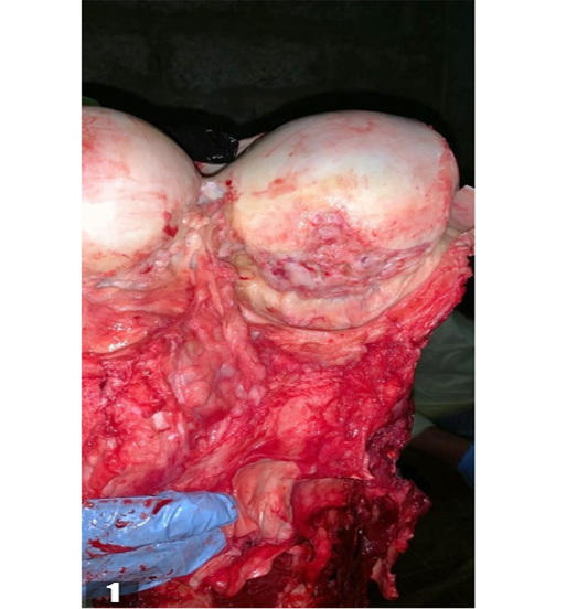
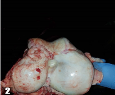
Figure 2: Distal end of Femur bone with synovial fluid having less viscosity and bone density was also found extremely low.

Figure 3: A chronic cracked sole at the junction of foot pad and skin penetrated deep into the food pad of right limb
Blood Chemistry
Blood sample of the elephant was taken for haematological analysis. Auricular or medial saphenous vein was the site of blood collection in elephant. Immediately after collection, the blood was transferred to 3 ml glass evacuated tube containing ethylenediaminetetraacetic acid (EDTA) for hematologic evaluations. The results of the haematological parameters of African elephant are presented in the Table 1.
Post-mortem Findings
Post- mortem examination was conducted under the supervision of chief veterinarian of the zoo. Bones, joints and joint cavities were examined. Articular surfaces were fragile and inflamed. Synovial Fluid was less viscous. Bone density was extremely low and mineralization of soft tissues were found on lateral articular surfaces of humerous and femur. (Figure 1 and 2 respectively). A cracked wound in the foot pad of right limb was observed. This chronic wound unbaled the elephant to walk again. Moreover this crackedsole penetrated the foot exposing the deep tissues to dirt and infection. (Figure 3). Therefore. It was tentatively diagnosed in necropsy report of animal that it was suffering from degenerative joint disease and aging factors. In another study, Chronic sole ulcerations linked with the degenerative joint disease in two Asian Elephants (Elephas maximus) was observed (Luikart and Stover, 2005). Similar results were found in the post-mortem examintation of 21 elephants (seven African Loxodonta africana and 14 Asian Elephas maximus), describing both pathology and variant anatomy. The most common pathological changes observed were bone remodelling, aging and Degenerative joint disease (Regnault et al., 2017).
Acknowledgements
We would like to acknowledge Riyadh Zoological Garden, Al Malaz Ar Riyadh, Saudi Arabia for facilitating required diagnostic and treatment facilities.
Conflict of Interest
All the authors promulgate no conflict of interest.
Authors Contribution
All the authors have contributed dedicatedly in terms of giving their technical expertise to give a tenable shape to this report.
References


