Advances in Animal and Veterinary Sciences
Research Article

A Study on Therapeutic Management, Prevention and Control of Contagious Pustular Dermatitis in Teddy Goat Herd in Pakistan
Nasir Iqbal1*, Ayesha Sadiq3, Zubair Luqman2, Hamza Jawad2, Sadaf Aslam1, Naveed Hussain1
1Department of Veterinary Surgery and Pet Sciences, University of Veterinary and Animal Sciences, Lahore, Punjab, Pakistan; 2Faculty of Veterinary and Animal Sciences, The Islamia University of Bahawalpur, Bahawalpur, Punjab, Pakistan; 3Department of Pathology, University of Veterinary and Animal Sciences, Lahore, Punjab, Pakistan.
Abstract | Contagious Pustular Dermatitis (CPD) or Scabby mouth is one of the most important diseases that mainly cause economic losses in goat sand sheep worldwide. This experimental trial is about the therapeutic management, prevention and control of Orf virus in teddy goat herd in Bahawalpur, Pakistan. Thirty two (32) teddy goats that were divided in two groups A and B having age range between 1-3 years were presented near Bahawalpur, Pakistan with history of anorexia, dullness and various types of cutaneous lesions around the oral cavity. The clinical picture was pointed toward Scabby mouth which is caused by Orf virus. Group A has been treated by Trisym (Symans Pharmaceuticals Private Limited) having trimethoprim 8% w/v and sulphadiazine 14% w/v and Melonac (ICI Pakistan Limited) having meloxicam 7.5mg/ml intramuscular for 5 days depicts better results than Group B has been treated by Trisym (Symans Pharmaceuticals Private Limited) having trimethoprim 8% w/v and sulphadiazine 14% w/v and Phlogen (ICI Pakistan Limited) having Flunixine meglumine 1mg/ml intramuscular for 5 days. Topically Somogel (Abbot Laboratories) having Lignocaine, eucalyptus, alcohol, menthol was applied in both groups over the lesions for 10 days after washing with potassium permanganate solution as an astringent. After 10 days of topical and 5 days of systemic therapy all goats were recovered. Glutaraldehyde results into better disinfectant as compare to virkon, 1% acetic acid and 2% sodium hypochlorite. Implementation of a contagious pustular dermatitis vaccination can be an important measure in economic losses reduction. By complete therapeutic treatment, disinfection of farm, proper vaccination and quarantine of the newly purchased animal the farm can lead towards decrease chances of contagious pustular dermatitis (CPD) and increase in milk and meat production.
Keywords | Teddy goat, Scabby mouth, Anorexia, Disinfection and necrotic lesions.
Received | May 31, 2020; Accepted | June 21, 2020; Published | July 28, 2020
*Correspondence | Nasir IQBAL, Department of Veterinary Surgery and Pet Sciences, University of Veterinary and Animal Sciences, Lahore, Punjab, Pakistan; Email: Nasir.iqbal@uvas.edu.pk
Citation | Iqbal N, Sadiq A, Luqman Z, Jawad H, Aslam S, Hussain N (2020). A study on therapeutic management, prevention and control of contagious pustular dermatitis in teddy goat herd in Pakistan. Adv. Anim. Vet. Sci. 8(9): 907-911.
DOI | http://dx.doi.org/10.17582/journal.aavs/2020/8.9.907.911
ISSN (Online) | 2307-8316; ISSN (Print) | 2309-3331
Copyright © 2020 Iqbal et al. This is an open access article distributed under the Creative Commons Attribution License, which permits unrestricted use, distribution, and reproduction in any medium, provided the original work is properly cited.
INTRODUCTION
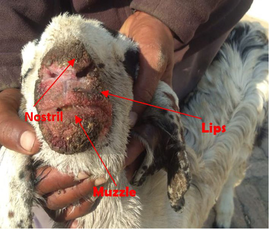
Contagious Pustular Dermatitis (CPD) is also known as Orf, contagious pustular dermatitis (CPD), sore mouth and contagious Ecthyma (de Oliveira, 2012). It is a contagious and zoonotic disease (communicable disease between humans and animals) that is caused by genus Parapoxvirus in the subfamily Chordopoxvirinae and family Poxviridae (Hosamani et al., 2007; Kumar et al., 2015). Parapox is dsDNA (double-stranded DNA virus) about 220-300nm long and 140-170nm wide with a unique property of an exclusive spiral coat that makes it distinguishable from other poxviruses (Hosamani, 2006; Jaisree et al., 2012). Chances of transmission are more in pasture feeding than the stall feeding because in pasture feeding animal is more exposed to trauma that may lead to scarified skin that mainly serves as a route of parapox transmission, results in the formation of macule, papule, vesicle, pustule and later that become a scab (Cargnelutti, 2011). Contagious pustular dermatitis has been distributed all over the world that raise goat, sheep, domesticated and wild ruminants (Adah et al., 2012). On clinical examination, there were ulcerated necrotic lesions around the muzzle, oral commissures and lips (de Oliveira, 2012). Rectal temperature, pulse rate and respiration were also elevated than the normal (Kumar et al., 2015).
Scabby mouth should be differentiated from some other abnormalities that are also involved in the development of lesions around the oral cavity like Peste des petits ruminant, Bluetongue, Goat pox and Foot and mouth disease but it does not involve digestive and respiratory system, does not involved tongue, do not have lesions other than face and it does not have lesions on other body parts so these diseases can be differentially diagnose from scabby mouth. It’s a viral disease. So, there is no specific treatment (McKeever et al., 1988). Supportive therapy and antibiotics has been given because animals become anorexic and more prone to secondary bacterial infection (Adah et al., 2012). Disinfection of animals can be done by using 1% acetic acid, Virkon, glutaraldehyde and 2% sodium hypochlorite by which disinfection of the farm can happen to avoid transmission of virus from herd (Gallina and Scagliarini, 2010). To control the scabby mouth animal must be isolated and quarantined before getting entry in the herd (Jaisree et al., 2012; Kumar et al, 2015).
MATERIALS AND METHOD
About thirty two (32) teddy breed goats herd was having scabby mouth were equally taken in two groups (A (n=16) and B (n=16)) for this experimental trial for a period of two weeks. All goats were from the same herd that was fed in pastures this has been depicted in flowchart.
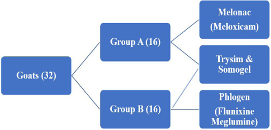
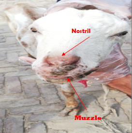
These 32 goats came with the history of anorexia, dullness and having dry ulcerated lesions around muzzle, oral commissures and lips from the last 4 days. On clinical examination, it was found that there are ulcerated necrotic lesions around the muzzle, oral commissures and lips (Figure 1). Rectal temperature, pulse rate and respiration were higher than the normal. There was no lesion on the coronet region. Conjunctiva was slightly pale. No sneezing, coughing or pneumonic sound was observed and no diarrhea was observed there. So, peculiar lesions and clinical pictures around the oral cavity pointed toward contagious pustular dermatitis (CPD) (Figure 2). For experimental therapeutic trial temperature, pulse and respiration of each animal in both groups was taken on day 1, 3, 5, 10 and 14. Group A (n=16) was treated with Trisym (Symans Pharmaceuticals Private Limited) having trimethoprim 8% w/v and sulphadiazine 14% w/v at a dose regime of 1ml/30kg was administered Intramuscular for 5 days along with Melonac (ICI Pakistan Limited) having meloxicam 7.5mg/ml at the dose rate of 1ml/33kg intramuscular for 5 days while Group B (n=16) was treated with Trisym (Symans Pharmaceuticals (Pvt.) Ltd.) having trimethoprim 8% w/v and sulphadiazine 14% w/v at a dose regime of 1ml/30kg was administered Intramuscular for 5 days along with Phlogen (ICI Pakistan Limited) having Flunixine meglumine 1mg/ml at the dose rate of 1ml/33kg intramuscular for 5 days. Topically Somogel (Abbot Laboratories (Pakistan) Limited) having lignocaine, eucalyptol, alcohol, menthol was applied on both groups over the lesions for 10 days after washing with potassium permanganate solution as an astringent. For the analysis of disinfection animals were divided in four Groups (A, B, C and D) having eight (8) animals in each group. Disinfection of Group A (n=8) can be done by using 1% acetic acid, disinfection of Group B (n=8) via Virkon and disinfection of Group C (n=8) by means of glutaraldehyde while disinfection of Group D (n=8) will be done by using 2% sodium hypochlorite. To control the transmission of virus isolation can be done for a period of 2 weeks.
Statistical analysis
Repeated measure ANOVA has been used to elaborate the findings.
RESULTS
Temperature, pulse and respiration of each animal in both groups was taken on day 1, 3, 5, 10 and 14. This has been shown in Table 1 revealing statistical analysis of Physical parameters (Temperature, Pulse, Respiration) of the animals between Group A and Group B at different intervals of days i.e. Day 1, Day 3, Day 5, Day 10 and Day 14 respectively. Group A animals with the treatment of Trysim and Melonac depicts marked decrease in temperature, pulse and respiration when compared with the treatment of Trysim and Phlogen. After 5 days of post therapy, the study shows that Meloxicam is far better than Flunixine meglumine when used for scabby mouth treatment.
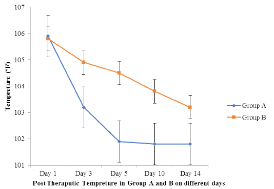
Figure 3 portray that by using Trysim and Melonac in group A there was a obvious decrease in temperature in animals as compare to the group B where treatment was done by using Trysim and Phlogen.
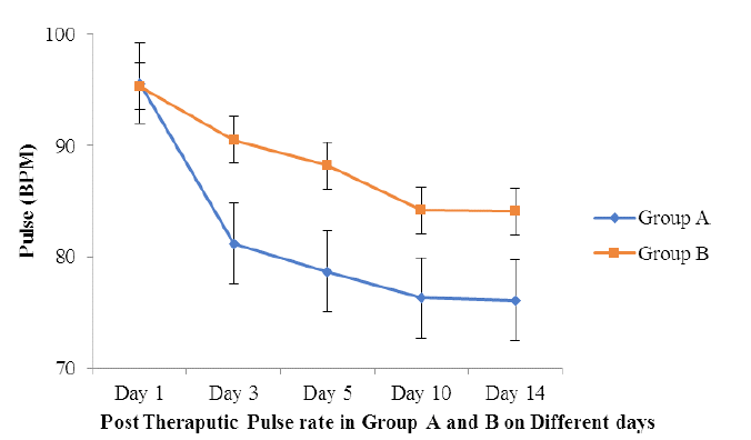
Figure 4 describes that by using Trysim and Melonac in group A pulse became normal within three days while in group B where treatment was done by using Trysim and Phlogen it happens after 10 days.
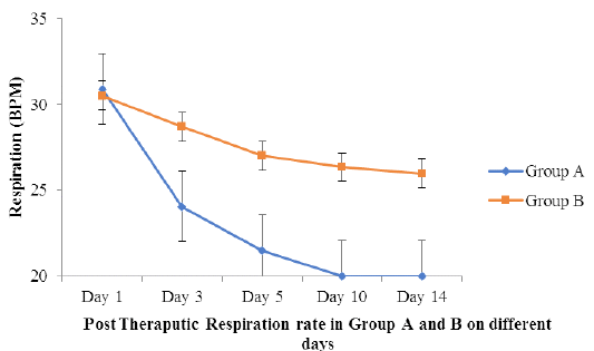
Figure 5 provides us information that group A have Trysim and Melonac treatment results into normal respiration rate in three to five days with a steep graph but in group B with Trysim and Phlogen treatment, respiration rate became normal after 10 days.
In Group A (n=16), Trisym (Symans Pharmaceuticals PVT LTD) having trimethoprim 8% w/v and sulphadiazine 14% w/v at a dose regime of 1ml/30kg was administered Intramuscular for 5 days along with Melonac (ICI Pakistan LTD) having meloxicam 7.5mg/ml at the dose rate of 1ml/33kg intramuscular for 5 days reveals better therapeutic management than Group B was treated with Trisym (Symans Pharmaceuticals PVT LTD) having trimethoprim 8% w/v and sulphadiazine 14% w/v at a dose regime of 1ml/30kg was administered Intramuscular
Table 1: Statistical Analysis of Physical parameters in Groups A and B on different days.
| Physical Parameters | Mean ±S.E. | Day 1 | Day 3 | Day 5 | Day 10 | Day 14 |
| Temperature | Group A | 105.9 ± 1.03 | 103.2 ± 0.99 | 101.9 ± 0.98 | 101.8 ± 0.98 | 101.8 ± 0.98 |
| Group B | 105.8 ± 1.06 | 104.9 ± 1.01 | 104.5 ± 0.99 | 103.8 ± 0.98 | 103.2 ± 0.98 | |
| Pulse | Group A | 95.6 ± 8.05 | 81.2 ± 7.52 | 78.7 ± 7.45 | 76.3 ± 7.42 | 76.1 ± 7.42 |
| Group B | 95.3 ± 8.12 | 90.5 ± 7.76 | 88.2 ± 7.53 | 84.2 ± 7.48 | 84.1 ± 7.45 | |
| Respiration | Group A | 30.9 ± 1.08 | 24.06 ± 0.99 | 21.50 ± 0.98 | 20.00 ± 0.98 | 20.00 ± 0.98 |
| Group B | 30.5 ± 1.32 | 28.69 ± 1.09 | 27.01 ± 1.03 | 26.35 ± 0.99 | 25.98 ± 0.99 |
for 5 days along with Phlogen (ICI Pakistan LTD) having Flunixine meglumine 1mg/ml at the dose rate of 1ml/33kg intramuscular for 5 days. Topically Somogel (Abbot Laborateries Pak LTD) having lignocaine, eucalyptol, alcohol, menthol was applied in both groups over the lesions for 10 days after washing with potassium permanganate solution as an astringent. Turpentine oil (t. t oil) was also applied there as a fly repellant. This has been depicted in Table 2 and Figure 6 as well.
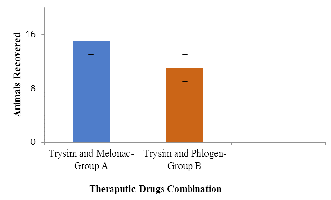
Table 2: Recovery of animals by using different therapeutic trials in Groups (A and B).
| Group no | Therapeutic drugs | Recovered animals |
| A (16) | Trisym, melonac and somogel | 15 |
| B (16) | Trisym, phlogen and somogel | 11 |
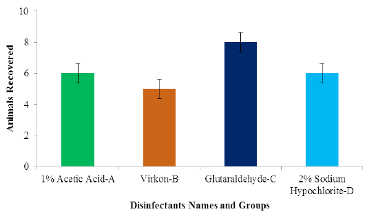
In all animals, the prognosis was good after 10 days of topical and 5 days of systemic therapy as all animals were recovered. For the analysis of disinfection animals were divided in four Groups (A, B, C and D) having eight (8) animals in each group. All disinfections portray satisfactory results but animals of Group C show better recovery as compare to others. This is also mentioned in Table 3. So, we can conclude that glutaraldehyde results into better disinfection than others.
Figure 7 reveals that by using glutaraldehyde disinfection can be better at farms as compare to the other disinfectants like virkon, 1% Acetic acid and 2% Sodium Hypochlorite. By isolation for a period of 2 weeks it also results into decrease chances of disease transmission as compare to the infected animals herding in normal animals.
Table 3: Recovery of animals by using different disinfectants in groups (A, B, C and D).
| Groups | Disinfectant name | Animals recovered |
| A | 1% Acetic Acid | 6 |
| B | Virkon | 5 |
| C | Glutaraldehyde | 8 |
| D | 2% Sodium Hypochlorite | 6 |
DISCUSSION
Scabby Mouth is not a lethal disease but from an economical point of view it is very important because the animal suffering from Scabby mouth results into decrease in milk production, increase treatment as well as farm disinfection cost by either chemical or heat treatment disinfection same as depicted by (Gallina and Scagliarini, 2010). After 10 days of topical and 5 days of systemic therapy all goats were recovered and the goats of Group A having Trysim and Melonac reveals better results than Group B having Trysim and Phlogen as it took 10 days to get animals normal. Implementation of a contagious pustular dermatitis vaccination in management schemes is still not widely applied by the farmers same as enlightened by (Savory et al., 2000). It can be an important measure that could greatly benefit farmers, as economic losses will be reduced along with improved production and minimizing greatly the impact of the disease also illustrated by (Spyrou and Valiakos, 2015). Group C having Glutaraldehyde as a disinfectant shows better results than Virkon, 1% Acetic Acid and 2% Sodium Hypochlorite but by using these disinfectants farm premises can be secure from this virus as Orf virus can be killed by this disinfection same as by (Gallina and Scagliarini, 2010). Newly purchased animals must be quarantine for about 14 days along with proper vaccination. This practice will result into less spread of this contagious disease similar as explained by (McKeever et al, 1988).
CONCLUSION
Contagious pustular dermatitis is not a fatal disease but is contagious which has a great impact on economy of the farmers especially in endemic areas, where the disease has spread in a great extent. It can be overcome by using trimethoprim and sulphadiazine along with meloxicam for 5 days. Topically oral wash with potassium permanganate solution and Somogel application over the lesions for 10 days will reduce the lesions. The prognosis is good as no major organ is affected in goats that were recovered. Proper disinfection by using glutaraldehyde, this virus can be prevented from the farm. Quarantine of animals before entry for two weeks in the herd can control the spread of the disease.
Acknowledgment
We want to acknowledge Dr. Naveed Hussain for his kind help.
Authors Contribution
Nasir Iqbal, Sadaf Aslam and Naveed Hussain: Research Trial and Revision.
Ayesha Sadiq, Zubair Luqman and Hamza Jawad: Formatting and Setting.
Conflict of Interest
The authors have declared no conflict of interest.
REFERENCES


