Advances in Animal and Veterinary Sciences
Research Article

Description of Hematological Value on Leopard Gecko (Eublepharis macularius)
Try Ardhi Wiranata1, Nove Hidajati2, Djoko Poetranto3, M. Gandul Atik Yuliani2, Retno Sri Wahjuni2, Widjiati Widjiati4*
1Post graduate Faculty of Veterinary Medicine Universitas Airlangga; 2Department of Basic Veterinary Medicine Science Faculty of Veterinary Medicine Universitas Airlangga; 3Department of Clinic Veterinary Faculty of Veterinary Medicine Universitas Airlangga; 4Department of Anatomy Veterinary Faculty of Veterinary Medicine Universitas Airlangga Jl. Mulyorejo, Surabaya - 60115, Jawa Timur, Indonesia.
Abstract | The aim of this research was to explore the hematological value of Leopard Gecko (Eublepharis macularius). Total of 20 samples were examined, it consists of 10 males and 10 females, blood samples were taken from vena ventral abdominal and deposited into ethylenediaminetetraacetic acid (EDTA) tubes, fresh blood leaves little to make blood smear, total blood examination and total leukocyte and differential leukocyte count used standard manual method. The mean result of hematological value of leopard gecko in this research were white blood cell (WBC) (x103/mm3) 7.12 male, 8.09 female, Lymphocyte (%) 46.2 male, 38.4 female, Monocyte (%) 17 male, 17.3 female, Heterophil (%) 18.3 male, 21.8 female, Eosinophil (%) 10.8 male, 13.9 female, Basofil (%) 7.7 male, 8.6 female, red blood cell (RBC) (106/mm3) 0.64 male, 0.65 female, Hemoglobin (Hgb) (g/dl) 8.95 male, 9.46 female, Hematocrit (%) 10.55 male and 9.26 female. In all parameters showed that hematology value of male gecko leopard was lower than female gecko except on two parameters which are hematocrit and lymphocyte which showed male gecko has the higher than females.
Keywords | Eublepharis macularius, Hematological, Total blood, Leopard Gecko, Description
Received | May 10, 2020; Accepted | June 22, 2020; Published | July 20, 2020
*Correspondence | Widjiati Widjiati, Departement of Anatomy Veterinary Faculty of Veterinary Medicine Universitas Airlangga, Email: widjiati@fkh.unair.ac.id
Citation | Wiranata TA, Hidajati N, Poetranto D, Yuliani MGA, Wahjuni RS, Widjiati W (2020). Description of hematological value on leopard gecko (Eublepharis macularius). Adv. Anim. Vet. Sci. 8(8): 882-887.
DOI | http://dx.doi.org/10.17582/journal.aavs/2020/8.8.882.887
ISSN (Online) | 2307-8316; ISSN (Print) | 2309-3331
Copyright © 2020 Wiranata et al. This is an open access article distributed under the Creative Commons Attribution License, which permits unrestricted use, distribution, and reproduction in any medium, provided the original work is properly cited.
INTRODUCTION
The Eublepharidae family currently has 36 different species and one of the most popular of all of these species is Eublepharis macularius or leopard gecko, according to Rawat et al. (2019) reported that leopard geckos are a very popular pet animal. Most pet store stock is believed to be captive bred at this time, with a mainly Pakistani ancestry. There are many color variations and they are great beginner pets. The reason why this species was a good pet for beginners because the character tended to calm and easy to maintain.
Leopard gecko has a commercial value and has been widely bred and traded freely to kept as a pet, accordance with the opinion of Landová et al. (2013) which states that the most favored lizard species by reptile hobbyists is leopard gecko (Eublepharis macularius), captivity has produced quite a lot of leopard gecko to complete the needs of the leopard gecko’s seekers, and thousands of these species are sold annually, rarely wild caught specimens are imported.
As the other pets, leopard gecko should be maintained and routinely checked for their health according problems in reptile health and welfare because owners simply do not know how to care for their pets Leopard geckos (Eublepharis macularius) are the second most common species of reptile (13% of all reptile cases seen) seen in private practice. The basic parameters used for the evaluation of reptile health are complete blood count (Perez, 2012).
Meanwhile, the value of hematological parameters on this species that reported are still rare, besides hematological parameters are widely used tools that aid in monitoring animal health, reproductive status, disease status and in the differentiation of physiological processes (Dissayanake et al., 2017). Values that are important to know for blood tests in reptiles are the total of erythrocytes, leucocytes, hematocrit, hemoglobin, and differential leucocytes (Stacy et al., 2015). Erythrocyte parameter in blood test used to evaluate general health, hydration and anemia also were observed in reptile patient (Stahl, 2006). Leucocyte parameter in blood test was important to help diagnose while reptile patient has viral, bacterial, fungal or parasites disease (Joseph, 2015). Based on the explanation, the author needs to do the observation for knowing the descrpition of total erythrocyte, leukocytes, hematocrit, hemoglobin, and differential leukocytes in the leopard gecko.
Materials and Methods
Materials
This research received ethical clearance released by Animal Care and Use Committee, Universitas Airlangga, Faculty of Veterinary Medicine (Number: 1.KE. 167.08.2019). Blood sampling was taken at the breeder and hobbyist of leopard gecko in Surabaya. Hematology values checkup inspection is in the Clinical Pathology laboratory, Faculty of Veterinary Medicine Universitas Airlangga, this research was a laboratory exploratory research with the design used is complete randomized sampling using 2 treatment groups and 10 replicates in each male and female treatment group. The data presented as means standard error of the mean (S.E.M) and were compared using the independent-samples T Test. They were considered to be significantly different if P < 0.05. The data obtained in this research is processed using SPSS program version 17.0.
Methods
Adult leopard gecko used as experimental animals was confirmed gender, all of the animal in a good condition and health, in this research using 10 male geckos and 10 females. Confirmed age was 9-18 months, birth data obtained from recording breeder. The length of the leopard gecko is sure to reach 15 - 25 cm using a ruler. The weight of gecko measured using scales, 60-90 g for male, and ≥45 g for female (Donoghue, 2016).
Before the blood sampling, rubbed the stomach using alcohol 70% to be sterile. Blood sampling was taken through the vena central abdominal (Perez, 2012). This vein branches in the umbilicus, therefore the needle is inserted into the cranial of the umbilicus (Olayemi, 2011). The blood sampling was taken using 1 ml disposable syringe with the 26 ½ g needle (Perez, 2012; Kobolkuti, 2009). Blood added slowly as much as 0,5 ml then syringe needle removed, and move the blood sample into the venoject which already given anticoagulant ethylenediaminetetraacetic acid (EDTA). Next check it with automatic hematology analyzer for total erythrocyte, hemoglobin, and hematocrit parameter and manual standard inspection for total leukocyte and leukocyte differential counting. The standard dilution of blood for leukocyte counts was 1:20; therefore, the dilution factor was 20. The volume of diluted blood used is based on the area and depth of the counting area. The area counted is 4 mm2 and the depth is 0.1 mm; therefore, the volume factor is 0.4 mm3. Total number of cells counted x dilution factor x 1/volume factor = cells/mm3. Average leukocyte counts from the duplicate pipets and report result (x 109/L or /mm3).
Protocol for blood staining
Giemsa’s solution was a mixture of methylene blue, eosin, and Azure B. A thin film of the specimen on a microscope slide were fixed in pure methanol for 30 seconds, by immersing it or by putting a few drops of methanol on the slide. The slide were immersed in a freshly prepared 5% Giemsa stain solution for 20–30 minutes (in emergencies 5–10 minutes in 10% solution can be used), then flushed with tap water and left to dry.
RESULT
The results of leukocyte count in this research were 9.7 x 10³/mm3 which was the highest value and the lowest value was 5.35 x 10³/mm3 with an average 7.12 x 10³/ mm3 in male geckos. In female geckos, the highest value was 9.4 x 10³/mm3 and the lowest value was 6 x 10³/mm3 with an average 8.09 x 10³/mm3. By using independent-sample t test with a significance level of 5% it can be concluded that the mean of male leopard geckosleukocyte is lower than female leopard gecko but the difference is not significant (p > 0.05) (Table 1).
The results of erythrocyte count in this research were 0.74 x 10⁶/mm3 which was the highest value and the lowest value was 0.37 x 10⁶/mm3 with an average 0.641 x 10⁶/mm3 in male geckos. In female geckos, the highest value was 0.89 x 10⁶/mm3 and the lowest value was 0.43 x 10⁶/mm3 with an average 0.651 x 10⁶/mm3. By using independent-sample t test with a significance level of 5% it can be concluded that the mean of male leopard geckos erythrocyte was lower than female leopard gecko but the difference was not significant (p> 0.05) (Table 1).
The results of hemoglobin count in this research are 10.5 g/dL which was the highest value and the lowest value was 5.3 g/dL with an average 8.95 g/dL in male geckos. In female geckos, the highest value was 11.6 g/dL and the lowest value was 8.1 g/dL with an average 9.46 g/dL. By using independent-sample t test with a significance level of 5% it can be concluded that the mean of male leopard geckos hemoglobin was lower than female leopard gecko but the difference was not significant (p > 0.05) (Table 1).
Table 1: Blood measurement in independent-samples T test.
| Type of blood |
Female Mean ± SD |
Male Mean ± SD |
Probability |
| Leukocyte | 8.09 ± 1.23 | 7.12 ± 1.40 | 0.680 |
| Erythrocytes | 0.65 ± 0.11 | 0.64 ± 0.10 | 0.903 |
| Hemoglobin | 9.46 ± 1,02 | 8.95 ± 1.37 | 0.833 |
| Hematocrit | 9.26 ± 2.18 | 10.55 ± 2.57 | 0.216 |
| Basophil | 8.60 ± 8.43 | 7.70 ± 5.45 | 0.069 |
| Eosinophil | 13.90 ± 7.57 | 10.80 ± 5.90 | 0.368 |
| Heterophil | 21.80 ± 5.92 | 18.30 ± 7.49 | 0.402 |
| Lymphocyte | 38.40 ± 11.07 | 46.20 ± 8.43 | 0.361 |
| Monocyte | 17.30 ± 5.83 | 17.00 ± 6.07 | 0.759 |
Note : no difference in the same column(p>0.05).
The results of hematocrit count in this research are 13.3% which was the highest value and the lowest value was 6.9% with an average 10.55%in male geckos. In female geckos, the highest value was 12.4 % and the lowest value is 6.1% with an average 9.26%. By using independent-sample t test with a significance level of 5% it could be concluded that the mean of male leopard geckos hematocrit was higher than female leopard gecko but the difference was not significant (p > 0.05) (Table 1).
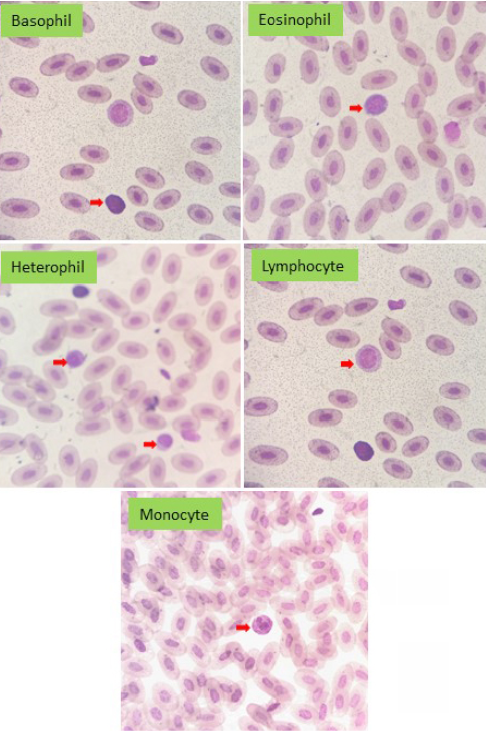
The results of basophil count in this research were 18% (1.22 x 10³/mm3) which was the highest value and the lowest value was 1% (0.05 x 10³/mm3) with an average 7.7% (0.57 x x 103/mm3) in male geckos. In female geckos, the highest value was 24% (2.26 x 10³/mm3) and the lowest value was 0% (0x 103/mm3) with an average 8.6% (0,69 x 10³/mm3). By using independent-sample t test with a significance level of 5% it could be concluded that in absolute value the mean of male leopard geckos basophil was lower than female leopard gecko but the difference was not significant (p > 0.05) (Table 1) and Figure 1.
The results of eosinophil count in this research were 24% (1.92x 10³/mm3) which was the highest value and the lowest value was 5% (0.32x 10³/mm3) with an average 10.8% (0.81x 10³/mm3) in male geckos. In female geckos, the highest value was 25% (1.95x 10³/mm3) and the lowest value was 2% (0.15x 10³/mm3) with an average 13.9% (1.12 x 10³/mm3). By using independent-sample t test with a significance level of 5% it could be concluded that in absolute value the mean of male leopard geckos eosinophil was lower than female leopard gecko but the difference is
not significant (p > 0.05) (Table 1).
The results of heterophil count in this research were 30% (2.10 x 10³/mm3) which was the highest value and the lowest value was 8% (0.54 x 10³/mm3) with an average 18.3% (1.27 x 10³/mm3) in male geckos. In female geckos, the highest value was 31% (2.73 x 10³/mm3) and the lowest value was 14% (1.08 x 10³/mm3) with an average 21.8% (1.75 x 10³/mm3). By using independent-sample t test with a significance level of 5% it could be concluded that in absolute value the mean of male leopard geckos Heterophil was lower than female leopard gecko and the difference is significant (p> 0.05) (Table 1) and Figure 1.
The results of lymphocyte count in this research were 60% (460x 103/mm3) which is the highest value and the lowest value was 36% (2.25 x 10³/mm3) with an average 46.2% (3.25x 10³/mm3) in male geckos. In female geckos, the highest value is 58% (5.39x 10³/mm3) and the lowest value was 27% (1.67x 10³/mm3) with an average 38.4% (3,14x 10³/mm3). One example of lymphocyte images that can be seen and entered in the calculations as in 4.12. By using independent-sample t test with a significance level of 5% it could be concluded that in absolute value the mean of male leopard geckos lymphocyte was higher than female leopard gecko but the difference is not significant (p > 0.05) (Table 1) and Figure 1.
The results of monocyte count in this research were 26% (2.13x 10³/mm3) which is the highest value and the lowest value was 9% (0.70x 10³/mm3) with an average 17% (1.19x 10³/mm3) in male geckos. In female geckos, the highest value was 25% (2.16x 10³/mm3) and the lowest value was 8% (0.60x 10³/mm3) with an average 17.3% (1.40x 10³/mm3). By using independent-sample t test with a significance level of 5% it could be concluded that in absolute value the mean of male leopard geckos monocyte was lower than female leopard gecko but the difference was not significant (p > 0.05) (Table 1) and Figure 1.
DISCUSSION
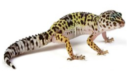
Eublepharis macularius or leopard gecko is anocturnal animal, it could be more active at night, living on land and can be found in desert areas such as Afghanistan, Pakistan, Mojave Desert, and West India (Figure 2). Leopard gecko is the most popular lizard among other types of lizards because its easy maintenance technique makes it become an ideal starts for beginners to pet the reptiles (Catchpole, 2012).
The taxonomic classification of leopard gecko is as follows: Kingdom: Animalia, Filum : Chordata, Class: Sauropsida, Ordo: Squamata, Family: Eublepharidae, Genus: Eublepharis, Species: Eublepharis macularius (Khan, 2009). Reference ranges for complete blood counts for a variety of reptile species have been published (Sykes and Klaphake, 2008). However, clinicians must try not to rely on these reference ranges as absolutes for the reasons described earlier. For many species of reptiles, there are no published normal values. Clinicians must rely on general trends and obvious changes in the haematological picture of the reptile patient (Stahl, 2006).
The results of leukocyte count in this research are 9.7 x 10³/mm3 which was the highest value and the lowest value is 5.35 x 10³/mm3 with an average 7.12 x 10³/ mm3 in male geckos. In female geckos, the highest value is 9.4 x 10³/mm3 and the lowest value is 6 x 10³/mm3 with an average 8,09 x 10³/mm3. Similar research by Olayemi (2011) mentions that leukocyte count of the females were slightly higher than males (1.563±3181.85 and 1.294±4067.68). Increases in leukocytes count are due to the increased physiological response of the body to respond acute infections/ inflammation as well as stressful conditions at the time of blood collection. While the decrease in the number of leukocytes can be caused by conditions that can damage the bone marrow, so that the bone marrow cannot make blood cells back, but can also occur because of damage to blood-forming agents caused by physical and chemical agents (Bijanti et al., 2010).
The results of the lymphocyte count in this research are 60% which was the highest value and the lowest value is 36% with an average 46.2% in male geckos. In female geckos, the highest value is 58% and the lowest value is 27% with an average 38.4%. Increased activity of the lymphocyte were associated with viral diseases, parasites, wound healing, ecdysis, immune stimulation. Decreases of the heterophils are associated with bacterial diseases, chronic stress, chronic malnutrition, immune suppression. Lymphocytes show reactivity by developing a basophilic cytoplasm or forming scalloped edges (Joseph, 2015).
The results of the monocyte count in this research were 26% which is the highest value and the lowest value was 9% with an average 17% in male geckos. In female geckos, the highest value is 25% and the lowest value is 8% with an average 17.3%. Increases of the monocyte are associated with chronic bacterial infection, granulomatous disease, parasites and chronic inflammatory disease. Monocytes become larger with exaggerated foamy cytoplasm. Cytoplasmic vacuoles and blebbing may occur with reactivity. A monocytosis suggests chronic or granulomatous inflammation. Monocytes have phagocytic capabilities and often engulf leukocytes and erythrocytes in response to anemia and infectious disease. They also actively engulf bacteria in a sepsis condition (Lim et al., 2017).
The results of the heterophil count in this research are 30% which is the highest value and the lowest value is 8% with an average 18,3% in male geckos. In female geckos, the highest value is 31% and the lowest value is 14% with an average 21.8%. Significant increases of the heterophils are associated with stress, neoplasia, chronic inflammatory diseases, bacteria and parasitic infection. Decreases of the heterophils are associated with viral infection and overwhelming septicemia. Heterophils become active and toxic with systemic illness. The severity of the systemic illness affects the degree of toxic changes present and the number of immature heterophils released into the peripheral blood. The toxic changes seen in the heterophil include an increase of cytoplasmic basophilia, degranulation, abnormal granulation (rounding), and vacuolization. The immature heterophils consist of bands, metamyelocytes and myelocytes (Joseph, 2015).
The results of the eosinophil count in this research are 24% which is the highest value and the lowest value is 5% with an average 10.8% in male geckos. In female geckos, the highest value is 25% and the lowest value is 2% with an average 13.9%. Eosinophil function in reptiles has not been well studied, abnormally high eosinophil numbers have been associated with parasitic infections, allergic reaction and immune stimulation, decreases the eosinophil significance not known (Stacy et al., 2015; Joseph, 2015).
The results of the basophil count in this research are 18% which is the highest value and the lowest value is 1% with an average 7.7% in male geckos. In female geckos, the highest value is 24% and the lowest value is 0% with an average 8.6%. Increases of the basophils are associated with respiratory tract disease and parasites (Stone et al., 2011).
The results of the erythrocyte count in this research are 0.74 x 10⁶/mm3 which was the highest value and the lowest value was 0.37 x 10⁶/mm3 with an average 0.641 x 10⁶/mm3 in male geckos. In female geckos, the highest value was 0.89 x 10⁶/mm3 and the lowest value was 0.43 x 10⁶/mm3 with an average 0.651 x 10⁶/mm3. Increase in the amounts of erythrocytes (polycythemia) according to their relative pathophysiology and absolute polycythemia. Polycythemia is a relative volume of plasma volume but the total volume of normal erythrocytes, whereas primary absolute polycythemia occurs due to increased erythrocyte volume, for example in polycythemia where the growth of erythrocytes becomes uncontrolled and leads to the production of erythropoietin and tumors in the kidney. Reduced amounts of erythrocytes are caused by decreased blood volume, decreased hemoglobin and vasoconstriction to increase delivery of 02 to vital organs (Bijanti et al., 2010).
The results of the hemoglobin count in this research were 10.5 g/dL which was the highest value and the lowest value was 5.3 g/dL with an average 8.95 g/dL in male geckos. In female geckos, the highest value is 11.6 g/dL and the lowest value was 8.1 g/dL with an average 9.46 g/dL. In general, the increase in red blood cells will be followed by an increase in hemoglobin, an increase in hemoglobin can be caused by acute dehydration, polycythemia vera, erythropoietin, lung disease, are in the plain high cancer, kidney and liver. Low hemoglobin levels can be caused due to lack of vitamins, especially vitamin B12, iron deficiency, there are disruptions to the spleen and chronic diseases such as kidney infection (Smith and Tangpricha, 2015).
The results of the hematocrit count in this research were 13.3% which is the highest value and the lowest value was 6.9% with an average 10.55%in male geckos. In female geckos, the highest value was 12.4 % and the lowest value was 6.1% with an average 9.26%. The higher percentage of hematocrit means the blood concentration was getting thicker. This happens because of fluid infiltration outside the blood vessels while the amounts of solids remain, the blood becomes more viscous, but can also be caused by dehydration, lack of oxygen supplies due to high altitudes, congenital heart disease and increased erythrocytes. Under conditions of hematocrit decrease occurs in individuals who experience acute blood loss (sudden blood loss), decreased erythrocyte level, vitamin B12 deficiency, malnutrition and overhydration (O’Leary and Samman, 2010). Age and season have been reported to affect blood parameter (including red blood cell (RBC), white blood cells (WBC), and platelet count among others) in a variety of new world primate species. These parameters can vary through the annual cycle or even throughout the life of individuals (Olayemi, 2011).
Conclusion
In all parameters showed that hematology value of male gecko leopard was lower than female gecko except on two parameters which are hematocrit and lymphocyte which showed male gecko has higher value than females.
Acknowledgements
The authors express sincere thanks to the Dean Faculty of Veterinary Medicine, Supervisor and co-Supervisor for providing all necessary facilities and fund for conducting research work.
authors contri bution
All authors contributed equally to the manuscript.
Research funding
This work is carried out with the support of the Ministry of Research, Technology and Higher Education of the Republic of Indonesia.
Conflicts of interest
The authors declare no conflicts of interest.
References


