Advances in Animal and Veterinary Sciences
Research Article

Prevalence of Most Common Feline Genital Surgical Affections in Teaching Veterinary Hospital, Cairo University, Egypt and Different Pet Clinics
Haithem A. Farghali1*, Nasser A. Senna1, Marwa S. Khattab2, Reda K.I. Shalaby3
1Department of Surgery, Anesthesiology and Radiology, Faculty of Veterinary Medicine, Cairo University, Giza, Egypt; 2Department of Pathology, Faculty of Veterinary Medicine, Cairo University, Egypt; 3Tanta Pet Animals Clinic, Tanta, Egypt.
Abstract | The present study was conducted to ascertain the prevalence of most common feline genital surgical affections in a teaching veterinary hospital, Faculty of Veterinary Medicine, Cairo University, Egypt and different pet clinics at Tanta and Cairo governorates during a 3-years period from October 2016 to March 2019. A total of 11334 cats (included 6029 queens and 5305 tomcats) belonging to different ages and breeds were included in this study. Feline genital system surgical affections were registered for age, sex, breed, and the main complaint from their owners. A full data of each case was collected including case history, clinical examination radiographic, ultrasonographic and laboratory findings in addition to histopathological examinations according to the previously designed examination sheet. A total number of 1684 of cats suffered from female genital surgical affections; 53 tomcats suffered from male genital surgical affections. The prevalence of uterine and ovarian surgical affections was recorded in addition to male surgical affections in tomcats. Female genital surgical affections were the highest recorded problems among cats. The present data enable veterinarians in Egypt to ascertain their needs for diagnostic tools and surgical interference that must be present at any pet clinic.
Keywords | Feline, Female genital surgical affections, Male genital surgical affections, Egypt, Prevalence
Received | August 20, 2019; Accepted | January 10, 2020; Published | June 25, 2020
*Correspondence | Haithem A. Farghali, Department of Surgery, Anesthesiology and Radiology, Faculty of Veterinary Medicine, Cairo University, Giza, Egypt; Email: dr_haithem0@yahoo.com
Citation | Farghali HA, Senna NA, Khattab MS, Shalaby RKI (2020). Prevalence of most common feline genital surgical affections in teaching veterinary hospital, Cairo university, Egypt and different pet clinics. Adv. Anim. Vet. Sci. 8(7): 709-719.
DOI | http://dx.doi.org/10.17582/journal.aavs/2020/8.7.709.719
ISSN (Online) | 2307-8316; ISSN (Print) | 2309-3331
Copyright © 2020 Farghali et al. This is an open access article distributed under the Creative Commons Attribution License, which permits unrestricted use, distribution, and reproduction in any medium, provided the original work is properly cited.
Introduction
Female genital affections are commonly seen in cats. Lesions in the ovaries and uterus may seriously influence normal reproductive capacity of cats and may put at risk the general health of the patients (Tawfik et al., 2015).
Pyometra (PYO) is one of the most common reproductive disorders in the queen, which is characterized by accumulation of purulent or mucopurulent material within the uterine lumen with an open or closed cervix during the luteal phase (Stanley and Pacchiana, 2008). An association between pyometra and cystic endometrial hyperplasia (CEH) has been established. In addition, this condition has been also observed in queens treated with progestogens (Schlafer and Gifford, 2008). Recommended treatment for CEH-pyometra in queens is ovariohysterectomy (OHE) with antibiotic and fluid replacement (Verstegen et al., 2008).
Uterine prolapse is a rare obstetrical emergency that occurs in feline species (Deroy et al., 2015; Jutkowitz, 2005) which was reported in both primiparous and multiparous animals. It usually arises during or within 48 hours of normal parturition, prolonged parturition or abortion (Özyurtlu and Kaya, 2005) when the cervix is dilated.
Uterine torsion is defined as the twisting of a uterine horn or uterine body about the longitudinal axis (Stanley and Pacchiana, 2008). It has rarely been reported in domestic animals, including cats. Uterine torsion is often related to pregnancy, and such cases occur during mid to late gestation (Matsumoto et al., 2009). Although the etiology of uterine torsion is unknown, broad ligament stretching caused by previous pregnancies, increased physical activity, uterine wall weakness, fetal movement and rough handling have been suspected as potential causes.
Ovarian remnant syndrome (ORS), which is defined as functional ovarian tissue present in a previously ovariectomized patient. Three explanations have been suggested for the development of ORS in companion animals (Sontas et al., 2007). The first and the most accepted explanation is incomplete surgical removal of one or both ovaries as a surgical error. This surgical error may be due to a small abdominal wall incision which hindering the ligature of an ovary (especially right one according to its anatomical location) or miss-ligation of both ovaries (Kalina et al., 2017).
Anatomically, the right ovary and uterine horn are located in a more cranial position than the left ovary and uterine horn and the suspensory ligament is shorter, making that ovary more difficult to exteriorize, which predisposes the surgeon to leave the ovary during the surgery. Therefore, the right ovary is more frequently found to be the remnant in the dog and cat (Ball et al., 2010).
Dropping of some ovarian tissue into the abdomen during the surgery was suggested as a second cause of ORS. If apiece of ovarian tissue is accidentally dropped into the abdomen during the surgery, this tissue revascularizes with omentum or the serosa of abdominal viscera and begins to function like a normal ovary (England and White, 2016). The third and final explanation is the presence of an accessory ovary or of ovarian tissue (ectopic) that is localized in the broad ligament or proper ligament of the ovary (Feldman and Nelson, 2004; Sontas et al., 2007). An accessory ovary is defined as an extra ovary, which is located adjacently and may be connected to the normal gonad. An accessory ovary has been reported in queens (Sontas et al., 2007).
The incidence of dystocia in cats has been described to vary between 0.4% and 8.0% and reach even higher numbers in certain breed groups. Dystocia may be due to functional causes (uterine inertia) or obstructive causes (maternal, fetal or a combination). Uterine inertia is the most common cause of feline dystocia. Other common causes include fetal malpresentation or malformations (Strom and Frossling, 2009).
Ovarian cysts, commonly arising from mature or atretic follicles, fail to ovulate and persistently remain on the ovary, thereby inhibiting re-establishment of folliculogenesis and consequently rendering the queen sub fertile. Cystic ovarian disease could be diagnosed based on behavioral signs, ultrasonography and histopathology (Eissa et al., 2017). In the queen, follicular cysts that arise from mature or atretic follicles are reported to be most common type. Affected queens may be asymptomatic or may exhibit prolonged estrus if cells lining secrete estrogen (Keskin et al., 2009).
Feline ovary neoplasm is rare. This most likely reflects an intrinsic resistance of cat ovaries to tumor development. Tumors of the ovary are classified based on their cell of origin-epithelial, germ cell or sex cord- stromal. In cats, epithelial tumors, germ cell tumors and sex cord-stromal turn have been reported (Gelberg and Mcente, 1985).
Cryptorchidism is rarer in cats than it is in dogs. In one study, 1.9% of intact male cats were cryptorchid. Persians are predisposed (Wood and Elder, 2009). Normally the testicles are in the scrotum at birth. Most cryptorchid cats present with an inguinal testicle (Dähnert and Wolfgang, 2011). The unilateral cryptorchidism was more frequent than bilateral (Ronald, 2011). Variety of congenital disorders lead to testicular hypoplasia or hypogonadism; testicular hypoplasia is rarer in cats. These affections were recognized in young animals as they reach puberty Johnson (2014).
The main object of this study was to determine the prevalence of the most common feline genital surgical affections compared with other soft tissue surgical affection that were admitted to the teaching veterinary hospital, department of surgery, anesthesiology and radiology, Faculty of Veterinary Medicine, Cairo University, Egypt and different pet clinics.
Materials and Methods
Ethical approval
All procedures of the current study were approved by and in accordance with the rules of animal use and care ethical committee of Faculty of Veterinary Medicine, Cairo University, Egypt.
Study area
The queens and tomcats used for this study were those admitted to the teaching veterinary hospital, Faculty of Veterinary Medicine, Cairo University, Giza, Egypt and different pet clinics in Cairo and Tanta provinces, Egypt for three years’ study period from October 2016 until March 2019.
Animals in the study
A total of 11334 cats (included 6029 queens and 5305 tomcats) which belonging to different ages and breeds were included in this study. This number included 3218 soft tissues surgical affected cats (included 1831 queens and 1387 tomcats), 7444 apparently healthy cats (included 4035 queens and 3409 tomcats) which admitted either for vaccination or for general health checkup, and 672 with orthopedic problems (included 163 queens and 509 tomcats).
Clinical investigation
All the cases were firstly recorded in the registration book for date, age, sex, breed, and complaint of their owners. Full clinical examination of each cats was carried out including complete medical, vaccination, dietary, and environmental history according to previously designed examination sheet (Sheet, 1). Visual inspection, rectal temperature, pulse and respiration rates of each case were recorded. In addition, examination of different organs and body systems was performed (Rijnberk and Stokhof, 2009).
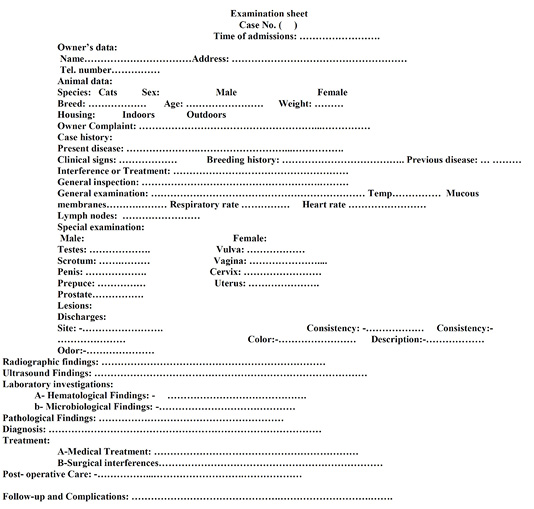
Sheet 1: Examination sheet.
Other diagnostic approaches
Radiological examination was applied according to (Parry and Lamb, 2010) using X-ray machine (Fischer imaging, with X- ray tube EMERALD-125, A-045211, Chicago, U.S.A.) and (EPSILON) 50-M, made in India.
Ultrasonography was applied according to (Kealy and McAllister, 2000) using ultrasonographic devices (TOSHIBA, 2004, made in JAPAN, CHISON, 2016, made in China and Mindray, 2010, CHINA).
Surgical techniques
Different surgical interventions included Castration which was achieved according to (Reichler, 2009). Cryptorchidectomy was applied by (Birchard and Nappier, 2008). All affected cats with cystic ovary, pyometra, uterine tumor, uterine torsion and uterine prolapse were treated by midline ovariohysterectomy (Fransson and Ragle, 2003), flank ovariohysterectomy (Coe et al., 2003) and Caesarian section (Traas, 2008).
All surgeries were performed under injectable general anesthesia. The cats were generally anaesthetized by (Atropine Sulphate, 0.05mg/kg) I/M or S/C and (Xylazine HCl, 2mg/kg) I/M as premedication. Then induction by (Ketamine HCl, 25-30 mg/kg) I/M and maintenance by Ketamine HCl were applied (Kaiser-Klinger, 2007). Another regime of injectable anesthesia for cats was by Atropine sulphate and Xylazine HCl as premedication. Then induction by (Propofol, Lipuro 1%) 1mg/kg I/V and maintenance by it (Vanlersberghe and Camu, 2008).
Histopathological examination
Histopathological examinations of different samples collected post-surgery in formalin saline were fixed in 10 % neutral formalin buffer. Fixed tissue was then processed by paraffin embedding technique. Tissue sections of 3-4 micron thick were made by microtome (Leica 2135, Germany). Tissue sections were then deparaffinized, rehydrated and stained by hematoxylin and eosin stain (Suvarna et al., 2012). Tissue sections were examined by light microscopy and photographed using camera Olympus XC30 (Tokyo, Japan).
Statistical analysis
Data that were collected about age, sex, breed, and season were collected and to Microsoft Excel 2010R spreadsheet, stored separately and exported to analytical software using Chi-square test. Values of < 0.05 were considered as statistically significant.
Result
In the present study, the total number of examined cats was 11334 cats (included 6029 queens and 5305 tomcats). Out of this number, 3218 were feline surgical soft tissues affections (included 1831 queens and 1387 tomcats), 7444 were apparently healthy cats (included 4035 queens and 3409 tomcats) which admitted either for vaccination or for general health checkup, and 672 were feline surgical orthopedic affections (included 163 queens and 509 tomcats), representing 28.4%, 65.6% and 6%, respectively. (Table1).
Table 1: The total number of examined cats and their Incidence.
| Queen | Tomcat | Total | Incidences | |
| Apparently healthy cat | 4035 | 3409 | 7444 | 65.6% |
| Soft tissues surgically cats | 1831 | 1387 | 3218 | 28.4% |
| Orthopedics surgically cats | 163 | 509 | 672 | 6% |
| total | 6029 | 5305 | 11334 | 100% |
The feline surgical soft tissues affections were recorded in 3218 cats among different systems. A total number of 1684 cats suffered from female genital surgical affections, 53 tomcats suffered from male genital surgical affections, 538 cats suffered from urinary surgical affections (included tom 462 cats and 76 queens), 392 cats suffered from different surgical skin affections (included 48 tomcats and 344 queens), 209 cats suffered from different surgical digestive affections (included 152 tomcats and 57 queens), 191 cats suffered from different surgical eye affections (included 117 tomcats and 74 queens), 89 cats suffered from different surgical abdominal wall affections (included 36 tomcats and 53 queens) and 62 cats suffered from different surgical ear affections (included 37 tomcats and 25 queens). The incidences of feline soft tissue surgical affections among different systems including genital system, urinary system, skin, digestive system, eye, abdominal wall, and ear were 53.9%, 16.7%, 12.2%, 6.5%, 5.9%, 2.8%, and 2%, respectively (Table 2).
Table 2: Number and incidences of feline soft tissue surgical affections among different systems.
| Different systems | No. of cases | Female | Male | % from total no of cases |
| Genital system | 1737 | 1684 | 53 | 53.9% |
| Urinary system | 538 | 76 | 462 | 16.7% |
| skin | 392 | 344 | 48 | 12.2% |
| Digestive system | 209 | 57 | 152 | 6.5% |
| Eye | 191 | 74 | 117 | 5.9% |
| Abdominal wall | 89 | 53 | 36 | 2.8% |
| Ear | 62 | 25 | 37 | 2% |
| Total | 3218 | 2313 | 905 | 100% |
From the obtained data, there were an eight most common surgical genital affections in examined queens including pyometra, Dystocia, Cystic endometritis, Ovarian cyst, Ovarian remnant syndrome, Uterine torsion, Uterine prolapse and Ovarian tumor which presented as 909, 468, 183, 94, 11, 8, 9 and 2 cases respectively with prevalence 54%, 27.7%, 10.8%, 5.6%, 0.6%, 0.5%, 0.57% and 0.11% out of total female genital affections respectively and 28.2%, 14.5%, 5.7%, 3%, 0.34%, 0.24%, 0.27% and 0.06% out of total soft tissue affections, respectively. (Table 3, 4 and 5).
The incidence of ovariohysterectomy in cats is recorded in pyometra, dystocia, cystic endometritis, ovarian cyst, uterine torsion, uterine prolapse and ovarian tumor at incidence 76.5 3.3, 10.3, 6.5, 1.4, 1.5 and 0.35%, respectively out of total female genital operations respectively (Figures 1-10) (Table 6).
The diagnosed surgical affections of the tomcats were Cryptorchidism and Testicular hypoplasia; this represented 1.58 % of total soft tissue affections (Table 7).
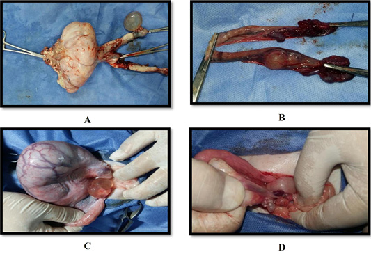
Figure 1: (A) Ovary of queen 12 years old suffered from cyst and large mass; (B) Ovary of queen 11 years old suffered from mass; (C) Ovary of queen 7 years old suffered from cyst; (D) Ovary of queen 5 years old suffered from many cyst.
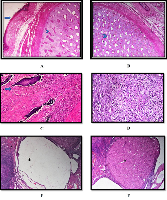
Figure 2: (A, B) Ovary of queen showing Ovarian in teratoma in which had well developed cartilage (arrowhead), and skin (arrow) (H and E stain 40); (C, D) Ovary of queen showing Severe neutrophilic in the interstitial which focal areas of liquefactive necrosis and calcification (suppurative oophoritis) (arrow) (H and E stain left X 200, right X400); (E, F) Ovary of queen showing follicular cyst (asterisk) (left) and luteal cyst (asterisk) (right) (H and E stain X100).
Table 3: Incidence of female genital affections.
| Affections | Total affected queens | % out of total female genital affections | % out of total soft tissue affections | Ages | Breeds | |||
| (1-2) years | Over 2 years | Mixed breed | Siamese | Persian | ||||
| Pyometra | 909 | 54% | 28.2% | 251 | 658 | 309 | 27 | 573 |
| Dystocia | 468 | 27.7% | 14.5% | 317 | 151 | 269 | 11 | 188 |
| Cystic endometritis | 183 | 10.8% | 5.7% | - | 183 | 69 | 7 | 107 |
| Ovarian cyst | 94 | 5.6% | 3% | - | 94 | 39 | 3 | 52 |
| Ovarian remnant syndrome | 11 | 0.6% | 0.34% | 11 | 2 | 9 | ||
| Uterine torsion | 8 | 0.5% | 0.24% | 6 | 2 | 1 | - | 7 |
| Uterine prolapse | 9 | 0.57% | 0.27% | 7 | 2 | 7 | - | 2 |
| Ovarian tumor | 2 | 0.11% | 0.06% | - | 2 | 1 | - | 1 |
| Total | 1684 | 100% | 45.9% | 840 | 489 | 697 | 48 | 939 |
Table 4: Incidence of different ovarian affections in queens.
| Affected organ | affections | No. | % out of total ovarian affections | % out of total female genital affections |
| Ovary | Ovarian cyst | 94 | 87.9 | 5.6 |
| Ovarian remnant syndrome | 11 | 10.2 | 0.6 | |
| Ovarian tumor | 2 | 1.9 | 0.12 | |
| Total | 107 | 100% | 6.3% |

Figure 3: (A) Radiograph for pyometra in queen showing a sausage-like fluid filled tubular organ; (B) B- mode ultrasound sagittal scan at the level of right flank region of a 3- years- old Persian cat suffered from closed pyometra. Notice hyperechoic thickened wall with intralumenal trapiculi and anechoic content of the right uterine horn.
Ultrasounds finding revealed hypoechoic testicle with hyperechoic rete testis at the mid of the testicle and the size was 1.8×0.6cm. The testicle located under the neck of the urinary bladder. In ectopic testicles homogenous hypoechoic testis with hyperechoic reta testis and testicular capsule were seen. The size of the affected testis was 1.8×0.9 cm (Figure 11).
Cryptorchidectomy gave satisfied results in two operated cats. Histopathological findings of ectopic testis showed that most seminiferous tubules composed of Sertoli cells only which separated from its basement membrane. Some seminiferous tubules had proliferation of its Sertoli cells lining indicating Sertoli cell tumor (Figure 11).
.
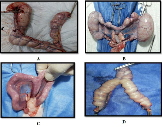
Figure 4: (A) Uterus of Persian queen 5 years old showing closed pyometra; (B) Uterus of mixed breed queen 8 years old showing closed pyometra; (C) Uterus of Persian queen 3 years old showing open pyometra; (D) Uterus of Persian queen 2 years old showing closed pyometra.
Discussion
Female genital affections in queens are rapidly becoming a major part of veterinary care. The present study indicated that uterine affections were the more frequent estimated affections than the ovarian ones. Some peoples prefer to raise female cats more than males (as it is evident in this work where queens were admitted about one and half times more than tomcats) and may be due to the disliked behavior of male cats during the mating season, such as spraying, which is not desired by many of the breeders.
Table 5: Incidence of uterine affections in queens.
| Affected organ | affections | No. | % out of total uterine affections | % out of total female genital affections |
| Uterus | Pyometra | 909 | 57.6% | 53.9% |
| Dystocia | 468 | 29.7% | 27.8% | |
| Cystic endometritis | 183 | 11.6% | 10.8% | |
| Uterine torsion | 8 | 0.5% | 0.47% | |
| Uterine prolapse | 9 | 0.6% | 0.53% | |
| Total | 1577 | 100% | 93.5% |
Table 6: Incidence of female genital system affections require OHE or Caesarian.
| Affections | Total affected queens | Operations | |
| OHE | Caesarian | ||
| Pyometra | 909 | 435 (76.5%) | - |
| Dystocia | 468 | 19 (3.3%) | 193 (100%) |
| Cystic endometritis | 183 | 59 (10.3%) | - |
| Ovarian cyst | 94 | 37 (6.5%) | - |
| Uterine torsion | 8 | 8 (1.4%) | - |
| Uterine prolapse | 9 | 9 (1.5%) | - |
| Ovarian tumor | 2 | 2 (0.35%) | - |
| Total | 1684 | 569 (100%) | 193 |
Higher incidence of feline genital system affections in Egypt rather than European and American countries, which may be regarded to routine spaying and castration in young age in these countries resulted in lowering the future affections of genital system (Stanley and Pacchiana, 2008; Strom and Frossling, 2009).
During this study, it was found that most cats who admitted to the veterinary hospital and clinics were for the follow-up routine (such as vaccination and deworming) and medical cases followed by soft tissue surgical affections and orthopedic affections representing 65.6%, 28.4% and 6% respectively out of total admitted cats. It was clear that more than one quarter of the presented cats were suffered from soft tissue surgical affections, which gave great attention to study such conditions.
As a side observation obtained from the present results, tomcats were frequently presented with surgical orthopedic affections more than female ones and it may be contributed to escape from homes during the mating season and exposure to falls from windows and road accidents.
Belonging to feline soft tissue surgical affections among different systems, the highest incidence was recorded in genital system followed by urinary system, skin, digestive system, eye, abdominal wall and ear with incidence percentage 53.9%, 16.7%, 12.2%, 6.5%, 5.9%, 2.8% and 2% respectively. The results, which indicates the importance of genital system affections studying.
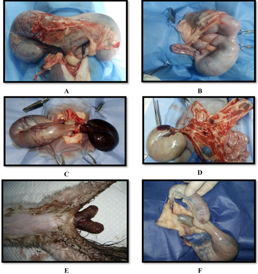
Figure 5: (A) Uterus of Persian queen 4 years old suffered from dystocia and subjected to OHE, note: presence of uterine adhesion at the level of uterine bifurcation; (B) Uterus of Persian queen 2 years old suffered from dystocia and subjected to OHE, note: presence of uterine adhesion at the level of right uterine horn; (C) Uterus of Persian queen 5 years old showing left uterine horn torsion, note: the torsion was at two levels, the first at the level of left ovary and the second at the level of uterine bifurcation; (D) Uterus of Persian queen 2 years old showing right uterine horn torsion, note: the torsion was at two levels, the first at the level of right uterine and the twisted part contained retained fetus; (E) Uterus of Persian queen one and half years old showing complete uterine prolapse; (F) Uterus of Persian queen 2 years old showing left uterine horn prolapse, note: the right uterine horn contained retained fetus.
The higher incidences of feline surgical affections among different systems were recorded in genital system which represent more than half the admitted soft tissue surgical cases (53.9%), followed by urinary, skin, digestive system, Sensory organs and abdominal wall with incidence percentage 16.7%, 12.2%, 6.5%, 7.9% and 2.8% respectively. The number of queens affected with genital surgical affections was 1684; this represented (45.9%) of total soft tissue affections. The affections included uterine and ovarian affections. The diagnosed surgical affections of the cat’s uterus and ovaries were pyometra, Cystic endometritis, dystocia, uterine prolapse, uterine torsion, ovarian tumor and ovarian cysts.
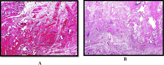
Figure 6: (A) Uterus of queen showing congestion, endema, hemorrhage, necrosis and few inflammatory cells infiltration in the endometrium (Uterine prolapse) (H and E stain X 100); (B) Uterus of queen showing endometrial necrosis and leukocytic infiltration in the case of Uterine torsion (H and E stain X 100).
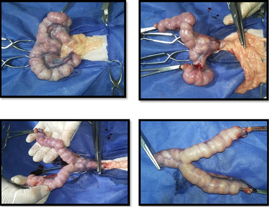
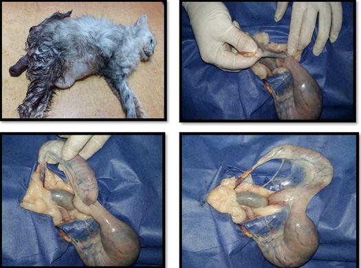

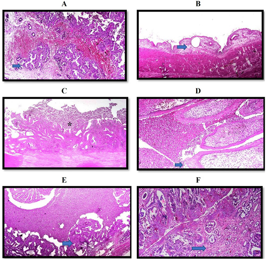
Figure 10: (A) Uterus of queen showing hemorrhage and Endometroid intraepithelial neoplasia Adenocarcinoma (arrow) (H and E stain X 100); (B) Uterus of queen showing Cystic endometrial hyperplasia (arrow) (H and E stain X 40); (C) Uterus of queen showing Cystic endometrial hyperplasia and pyometra. The uterine lumen is dilated and filled with neutrophils and necrotic debris (asterisk). The myometrium is thin and stretched. Some of the endometrial stroma contains mostly mononuclear cells (H and E stain X 40); (D) Cervix of queen showing papillomatosis, cytoplasmic clearing [Halos) of the epithelium (arrow) and necrotic debris and neutrophils in the lumen (H and E stain X 200); (E) Uterus of queen showing Cystic endometrial hyperplasia and pyometra in which the lumen is filled with neutrophils and necrotic (arrow) (H and E stain X 100); (F) Uterus of queen showing leukocytic infiltration in the endometrium which characterizes endometritis and mild peri-glandular fibrosis (arrow) (H and E stain X 100).
Table 7: Incidence of male genital affections in tomcat.
| Affections | No. | % out of total male genital affections | % out of total soft tissue surgery | Age | Breed | |||
| (6-24ms) | Ages Over 2years | Mixed breed | Siamese | Persian | ||||
| Cryptorchidism | 51 | 96.3% | 1.5% | 51 | - | 17 | 34 | |
| Testicular hypoplasia | 2 | 3.7% | 0.06% | 2 | - | - | 2 | |
| total | 53 | 100% | 1.56% | 53 | 17 | 36 | ||
Cats are generally considered induced ovulators and are subjected to multiple cycles of estrogenic stimulation. If a queen ovulates but does not conceive, she may undergo pseudocyesis and eventually develop pyometra or endometritis (Potter et al., 1991). The incidence of pyometra is lower in queens than in bitches, as queens are induced ovulators (Verstegen and Onclin, 2006).
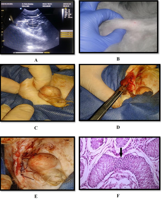
Figure 11: (A) B-mode sagittal scan in an 8 months old Persian tomcat suffered from unilateral ectopic testis. Notice the homogenous hypoechoic testis with hyperechoic reta testi and testicular capsule. The size of the affected testis was 1.8×0.9 cm; (B) Abdominal tests; (C) Ectopic testis; (D, E) Cryptorchidectomy; (F) Seminiferous tubules showing proliferation in Sertoli cells (Sertoli cell tumor) (arrow) (H & E×200).
Our study revealed that the most prevalent uterine affection in tested queens was pyometra at incidence 57.6 % out from total uterine affection and 53.9 % out from female genital affection followed by dystocia and cystic endometritis. On the contrary, (Scott et al., 2002; Stanley and Pacchiana, 2008) reported that Pyometra in cats is not common, 12 of three thousand (0.4%) adult free-roaming females were found having pyometra. These findings may be contributed to the fact that most of Egyptian cat owners did not sterilize their cats and in the same time they did not frequently breed them that gave the chance to hormonal predisposing factors of pyometra induction. In addition to owner-dependent widely misuse of contraceptive medications in Egypt.
Incidence of uterine torsion and prolapse are rare in present study, although (Silva, 2008) reported that Uterine torsion is more frequently described in cats. It may be due to that these affections were mainly associated with pregnancy and parturition which are less common within cat owner’s society in Egypt (Figures 5,6 and 8).
The most common clinical symptoms associated with pyometra were inappetence, with or without fever, depression, polydipsia, lethargy, with or without vaginal discharge and abdominal distension. Moreover, abdominal examination revealed distension and pain. Similar signs were recorded before (Debosschere et al., 2001; Agudelo, 2005; Smith, 2006).
Ultrasonography was a helpful tool for diagnosis and treatment of various surgical affections of genital tract (Abu-Seida and Torad, 2012). In the present study, pyometra had characteristic ultrasonographic features as fluid filled uterus with variable wall thickness and proliferative changes (Figure 3). Similar findings were also mentioned (Bigliardi et al., 2004; Parmigiani et al., 2004).
Our findings revealed that dystocia is more common in Persian cat than other breeds, this similar to in the cat, the incidence of dystocia is known to be highest in the Persian cat (Widmann Acanal, 1992; Ekstrand and Linde-Forsberg, 1994) followed by the Siamese type cats (Gunn Moore and Thrushfield, 1995).
In the present study, Cystic endometritis is a common in queen over two years; similar findings were record by (Thrall, 2004) reported that cystic endometritis common in queens older than three years and in other queens older than five years without relationship to the number of parturitions. On the other hand, the average age of queens suffering pyometra was seven years (Johnston et al., 2001). Cystic endometritis was more prevalent in small breeds than large ones, while the condition was less observed in queens presumably due to its induced ovulatory nature (Hedlund, 2002).
In the present study, ovarian cyst was the most common ovarian affections 87.9 % in queen followed by ORS (10.2%). The most common type of ovarian cyst is the functional follicular cyst, which secretes estrogen (Johnston et al., 2001). Follicular cysts could cause behavioral disorder like prolonged estrus, bone marrow suppression and uterine hyperplasia due to prolonged elevation of estradiol. Follicular cysts could be diagnosed based on behavioral signs, ultrasonography and evaluation of serum estradiol (Gelberg et al., 1984; Johnston et al., 2001).
Kalina et al. (2017) summed up that the ORS is a rare iatrogenic condition, especially in cats, caused not only by inexperience of the surgeon, in which are observed too variable signs of estrus under hormonal influence. They may appear even up to several years after OVH, but an average of about 8 months has been registered, as usually the syndrome combines with uterine stump pyometra.
From the obtained data, the most common surgical affections of uterus were pyometra representing 57.6% out of total uterine affections followed by dystocia representing 29.7 % out of total uterine affection. These results agree with (Senna et al., 2015) who reported similar incidence of pyometra (54.2%) in uterine affection of cats. On the contrary, these results disagree with (Scott et al., 20002; Stanley and Pacchiana, 2008) who published that pyometra in cats is not common (0.4%).
In the present study, prevalence of ovarian tumor in queen was 1.9%, these findings similar to (Miller et al., 2003) who reported in one study that uterine tumors are rare in cats; constituting 0.29% of feline neoplasms. In addition, (Cotchin and Marchant, 1977; Gelberg and Mcentee, 1985) reported that neoplasms arising in the feline ovary are rare. In cats with uterine tumors, the following clinical signs are presented; abdominal mass, weight loss, anorexia, pain, and vaginal bleeding in some cases (Cooper et al., 2006).
The obtained results proved that Ovariohysterectomy as a surgical interference is still the ideal approach for dealing with pyometra and cystic endometritis, while Caesarian ideal approach for dystocia. These results agree with those previously mentioned by (Bigliardi, 2004; Agudelo, 2005; Smith, 2006; Younis et al., 2014).
Cryptorchidism the most detected surgical affection in male genital systems followed by testicular hypoplasia. These results agreed with (Senna et al., 2015) reported that cryptorchidism was recorded in 70.6% from the total affected tomcats. On the contrary, these results disagree with (Scott et al., 2002; Ronald, 2011) who reported that cryptorchidism is a rare condition in cats, with incidence ranging from 0.4-2 %. In addition, the prevalence of cryptorchidism in cats has been reported as 1.3–3.8%, which is generally lower than the prevalence in dogs (Yates et al., 2003).
The high incidence of cryptorchidism may be due to inexpert selection of cat breeders of pure bloodline as (Johnston et al., 2001). Reif and Brodey (2005) said that it was congenital or inherited disease. The Persian breed cats showed a larger prevalence of cryptorchidism than other breeds, these results agree with (Millis et al., 1992) who found that Persians cats to be predisposed to cryptorchidism than other breeds.
Our findings revealed that pyometra and cryptorchidism were the most prevalent surgical affections in genital systems of queens and tomcats, respectively. These findings agreed with (Senna et al., 2015) reported that the most detected genital affections in queen and tomcats, respectively.
Conclusion
It was concluded that genital system affection the most prevalent affections among soft tissue surgical affections. Pyometra and dystocia was the most prevalent surgical affections among female genital system in queens, while Cryptorchidism followed by Testicular hypoplasia was the most prevalent surgical affections in male genital of tomcats. This study suggests that most cases of ovariohysterectomy are performed secondary to pyometra, cystic endometritis and ovarian cyst.
Acknowledgments
The research was approved by the scientific committee of Cairo University for animal research and animal welfare, and was carried out in teaching veterinary hospital, Cairo University and Tanta Pet Animals Clinic. The authors are indebted to the cat’s owners for their endless collaboration.
Authors Contribution
Haithem A. Farghali contributed to the conception and design of the work and surgical interventions in addition to writing of the manuscript. Nasser A. Senna contributed to the conception and design of the work in addition to revision of the manuscript. Marwa S. Khattab contributed to the histopathological examination and interpretations. Reda K.I. Shalaby contributed to the collection of the data, clinical examination and surgical interventions.
Conflict of interest
There is no conflict of interest between authors.
References


