Advances in Animal and Veterinary Sciences
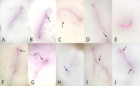
Effects of administration vanadium pentoxide on sperm morphology in rats. A) normal sperm, (B) coiled tail, (C) looped tail, (D) curved tail, (E) detached head, (F) abnormal head shape, (G) broken head, (H) cytoplasmic droplet, (I) detached head, and (J) straight head with cytoplasmic droplet (original magnification 100×; eosin and nigrosine stains).
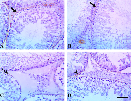
Testicular α-smooth-muscle actin (α-SMA) immunostaining in rats treated with quercetin and vanadium pentoxide (VP). Representative photomicrographs show changes in myoid cells (brown pigment) via α-SMA immunostaining of testicular sections in (A) control, (B) quercetin-treated, (C) VP-treated, and (D) quercetin and VP co-treated groups. The control and quercetin-treated groups showed positive, strong α-SMA immunostaining (solid arrows), whereas VP-treated rats showed weak α-SMA immunostaining (dashed arrow), and the quercetin and VP co-treated rats showed moderate α-SMA immunostaining (arrowhead). Scale bar: 50 µm.
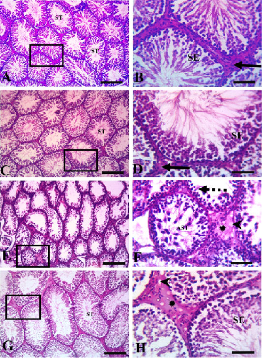
Testicular histopathology in rats treated with quercetin and vanadium pentoxide (VP). Photomicrographs of testicular sections. Control group (A &B), quercetin group (C&D), VP treated-group (E &F), quercetin +VP treated-group (G&H). B, D, F, H represents higher magnifications of the boxed area in A, C, E, and G, respectively. The control and quercetin-treated rats (A, B, C, and D) showed normal histological structures of closely packed seminiferous tubules (ST) lined with seminiferous epithelial cells (SE), and interstitial tissue containing Leydig cells (solid arrows). The VP-treated group (E and F) showed severe germinal epithelial atrophy (dashed arrows), with interstitial tissue replaced with homogenous eosinophilic materials (asterisk) and vacuoles (arrowheads). VP-induced abnormalities were less evident in the VP and quercetin co-treated group (G and H). Scale bars A,C, E; 200 µm , B,D,F 50 µm.


