Advances in Animal and Veterinary Sciences
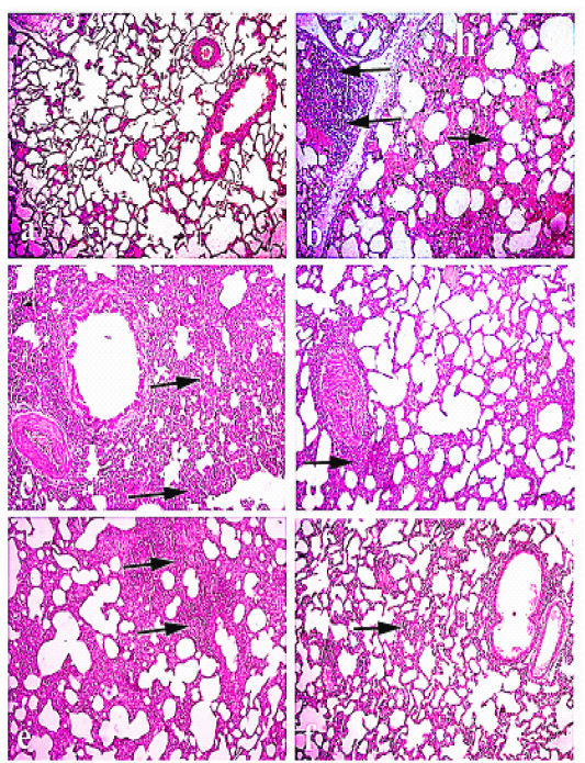
Lung of rabbit: (a) Lung of normal control animals. (b)and (C) lung of pasteurella infected group showing interstitial inflammatory reaction and hemorrhage, and (d) lung treated with Ofloxacin., (e)lung treated with Grape seed extract . (f ) lung treated with( O+ G). (arrows) refer to thickening of interstitial tissue with lymphocytic infiltration. Hematoxylin and eosin stain; magnification, 100×.
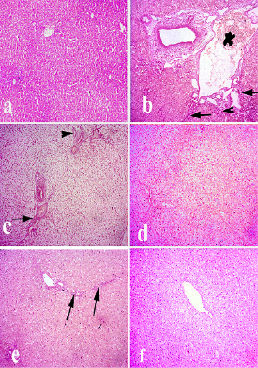
Liver of rabbit. (a) liver of control animals. (b and c) liver of pasteurella infected group showing degeneration. necrosis of hepatocytes and severe congestion of blood vessels, hyperplasia of bile duct and fibrosis. (d) livertreated with (Ofloxacin) showing diffuse vacuolar degeneration. (e) livertreated with grape seed extract showing mild vacuolar degeneration of hepatocytes along with fibrosis. (f) liver treated with (O+G) showing mild focal pericentral vacuolar degeneration. (arrows) refer to fibrosis and lymphocytic infiltrations, (arrow heads) refer to hyperplasia of bile ducts. Hematoxylin and eosin stain; magnification, 100×.
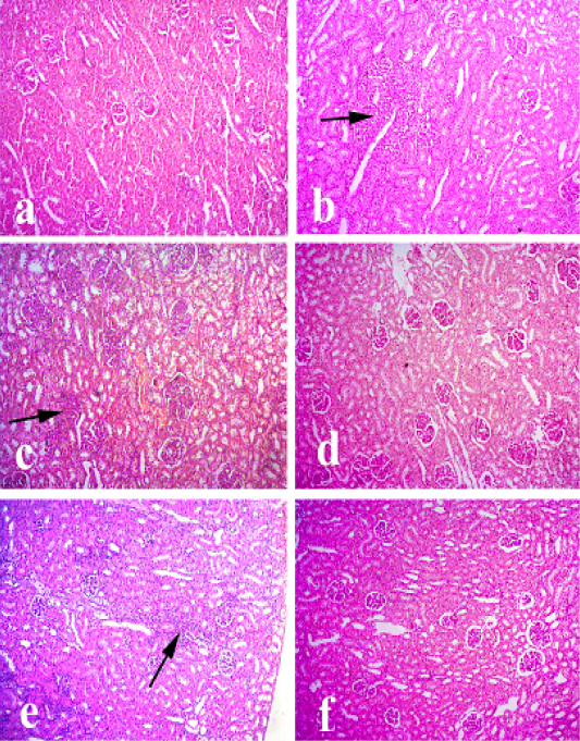
Kidney of rabbit. (a) Kidney of control animals. (b and c) Kidney of pasteurella infected group showing degeneration of renal tubules, proliferation of some glomeruli and focal interstitial aggregations with lymphocytes. (d) Kidney treated with (Ofloxacin) showing mild vacuolar degeneration of some renal tubules. (e) Kidney treated with grape seed extract showing mild vacuolar degeneration of some renal tubules along with mild lymphocytic infiltrations between the renal tubules. (f) Kidney treated with (O+G) showing pronounced improvement of renal tissue. (arrows) refer to lymphocytic infiltrations. Hematoxylin and eosin stain; magnification, 100×.
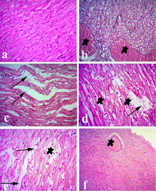
Heart of rabbit. (a) Heart of normal control animals showing normal heart muscels. (b and c) Heart of pasteurellainfeceted group showing severe congestion of blood vessels and interstitial inflammatory reaction, degeneration andintermuscular edema. (d) Heart treated withofloxacin showing mild congestion and mild degeneration and intermuscular edema. (e) Heart treated with grape seed extract showing mild congestion and mild degeneration and intermuscular edema. (f) Heart treated with (O+G) showing fairly normal histological picture. (arrows) refer to degeneration and intermuscular edema, (stars) refer to congestion. Hematoxylin and eosin stain; magnification, 100×.
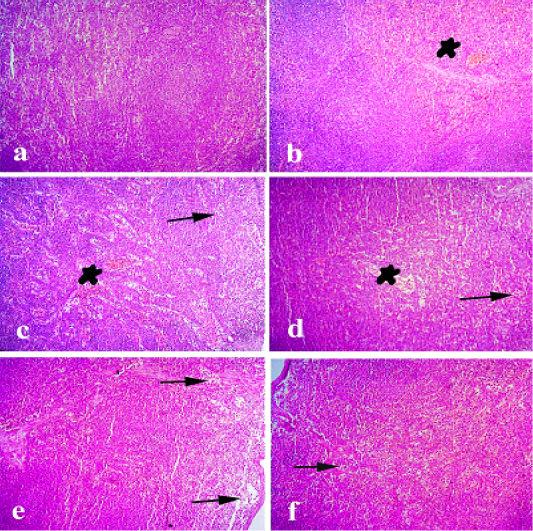
Spleen of rabbit. (a) Spleen of normal control animals showing normal white and red pulps. (b and c) Spleen of pasteurella infected group showing severe congestion and focal depletion of white pulp. (d) Spleen treated with ofloxacin showing mild congestion of sinusoidal blood vessels. (e) Spleen treated with grape seed extract showing mild depletion of white pulp. (f) Spleen treated with(O+G) showing discrete depletion of some lymphocytes. (arrows) refer to depletion of white pulp. (stars) refer to congestion of blood vessels. Hematoxylin and eosin stain; magnification, 100×.
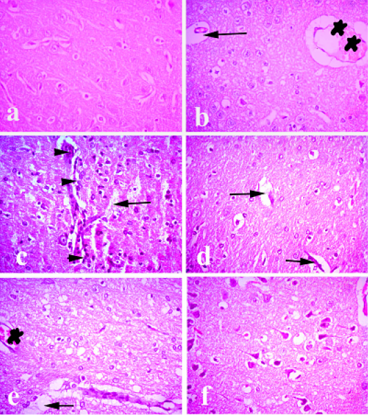
Brain of rabbit. (a) Brain of normal control animals showing normal histological picture. (b and c) Brain of pasteurella infected group showing congestion of blood vessels, lymphocytic infiltrations and degeneration of neurons. (d) Brain treated with ofloxacin showing focal degeneration of some neurons. (e) Brain treated with grape seed extract showing mild congestion and focal degeneration. (f) Brain treated with (O+G) showing fairly normal picture. (arrows) refer degenerated neurons, (arrow heads) refer to lymphocytic infiltration. (stars) refer to congestion. Hematoxylin and eosin stain; magnification, X 100.


