Advances in Animal and Veterinary Sciences
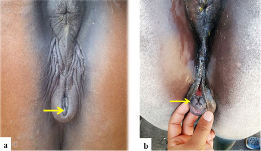
a and b Maiden mare affected with granulosa cell tumor showing well-developed clitoris.
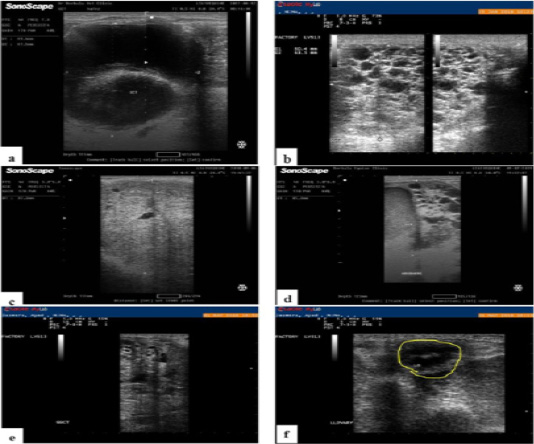
a, Ultrasound image of Arabian mare ovary with granulosa cell tumor showing double large cystic dilatations. b, Ultrasonographic image of Arabian mare ovary with GCT showing multiple cysts with typical honeycomb appearance. c, Ultrasound image of ovary of Arabian mare with granulosa cell tumor. Note the solid mass with few very small cystic cavitations. d, Ultrasound image of Arabian mare ovary with granulosa cell tumor. Note presence of mixed pattern of the tumor. e, ultrasonography showing right ovary with GCT and f, atrophied and non-functioning left ovary of the same case.
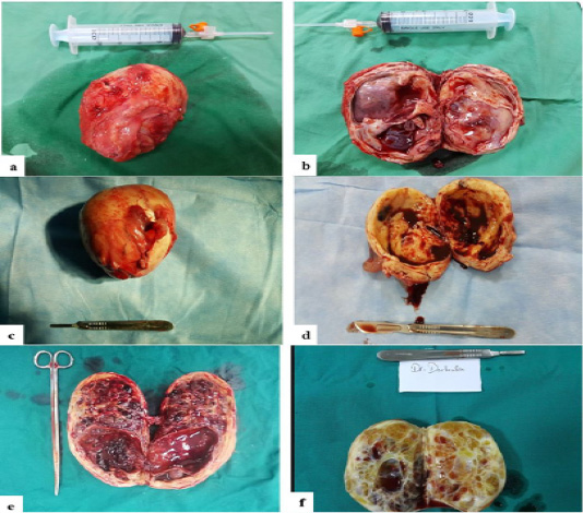
a, Gross appearance of surgically removed ovary of Arabian mare with granulosa cell tumor showing enlarged ovary. b, On section of the same ovary, the tumor reveals multiple large cysts with atrophied ovarian tissue. c, Arabian mare ovary with granulosa cell tumor showing ovary with increased size. d, On section, the tumor has a central large cavity filled with hemorrhagic fluid that exerts pressure atrophy on ovary. e, incised ovary of Arabian mare showing mixed type of granulosa cell tumor, the tumor reveals a large cystic cavity and multiple small cysts. f, a section of Arabian mare ovary. Note the thickened ovarian wall with multiple irregular small cystic spaces (honey comb- like appearance).
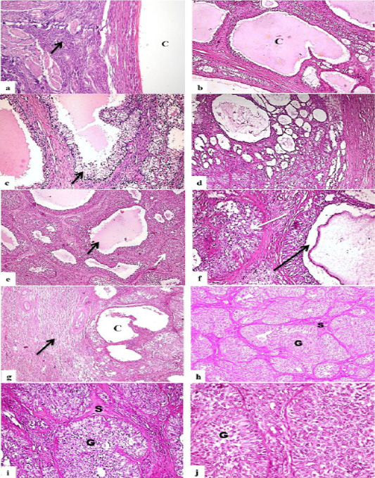
Photomicrograph, ovaries of Arabian mares with GCT. a, ovary showing single large cyst lined with granulosa cells (C), note remaining inactive ovarian tissue (arrow) (H and EX200). b, ovary showing variably-sized cysts (C) that lined with granulosa cells and their lumen filled with homogenous esinophilic structure (HandEX200). c, ovay with GCT, note the multiple large cavities that lined with granulosa cells (arrow) and filled with homogenous esinophilic structure (H and EX200). d, ovary showing multiple and variably-sized cystic cavitation in form of honey-comb like pattern (H and EX100). e, ovary showing a mixed pattern of follicular (black arrow) and solid mass (white arrow) GCT (H and EX200). f, higher magnification of the previous photo showing the follicular-like pattern (black arrow) and solid pattern (white arrow) (H and EX400). g, ovary with mixed solid and cystic GCT (C) showing inactive ovarian tissue surrounding focal tumor mass (arrow) (H and EX100). h, ovary showing solid granulosa cells tumor, tumor cells tend to be arranged in a group of cells (G), each group of cells was separated by connective tissue stroma (S) (H and EX100). I, solid GCT showing groups of granulosa cells (G) surrounded by fine connective tissue stroma (S) (H and EX200). j, higher magnification of the previous photo showing large, vacuolated granulosa cells with prominent nuclei (G) (H and EX400).


