Advances in Animal and Veterinary Sciences
Research Article
Dissociation of Mycobacterium bovis: Morphology, Biological Properties and Lipids
Olexiy Tkachenko1*, Maryna Bilan1, Volodymyr Hlebeniuk1, Natali Kozak1, Vitalii Nedosekov2, Olexandr Galatiuk3
1Dnipro State Agrarian and Economic University, Serhii Efremov Street, 25, Dnipro, 49600, Ukraine; 2National University of Bioresources and Management of Nature of Ukraine; 3Zhytomyr Agrarian and Ecological University, Ukraine.
Abstract | The article presents the results of investigations concerning the cleavage of avirulent cells from the virulent epizootic strain Mycobacterium bovis (M. bovis) during the 20 month storage at low positive temperatures (2-3°C). It was preceded by a 116-fold reinoculation of the suspension of the virulent culture into a dense nutrient medium. Mycobacteria of the detected colonies have acquired new features and properties. It was noted that in the second and subsequent generations, mycobacteria appeared in the form of non-acid resistant grains and short rod-shaped bacteria and L-forms of the protoplastic (spheroplastic) type. The change in morphology was accompanied by the emergence in mycobacteria of the ability to be grown on chemically simple nutrient media. Different morphological forms of mycobacteria were found in the elective dense nutrient medium at 3°C and 37°C cultivation: at 3°C irrespective of the initial morphological forms, non-acid-resistant short rod-shaped bacteria and grains were generated in dynamics; at 37°C – L-shaped protoplastic (spheroplastic) type and elementary small bodies. The appearance of the latter in culture has changed the nature of its growth. It is shown that qualitative processes of morphological forms of the biological cycle of mycobacteria occur on the background of quantitative changes in the chemical composition, in particular lipids. The content of total lipids decreased, at the stability of the quality of the fractions of total lipids. Differences in the ratio of unsaturated to saturated fatty acids in pathogenic and non-pathogenic mycobacteria have been established. Long-chain acids with a content of 26-27 carbon atoms, regardless of the temperature of cultivation, disappeared in the dissociative forms. The biological cycle of the development of dissociative M. bovis at different temperatures of cultivation with properties distinct from the parent strain, characterizes the wider possibilities of the investigated microorganism as it is known today.
Keywords | Mycobacterium bovis, Dissociative forms, Low-temperature conditions, Biological properties, Morphology, Lipids
Received | November 06, 2019; Accepted | January 02, 2020; Published | March 05, 2020
*Correspondence | Olexiy Tkachenko, Dnipro State Agrarian and Economic University, Serhii Efremov st., 25, Dnipro, 49600, Ukraine; Email: bilan.m.v@dsau.dp.ua
Citation | Tkachenko O, Bilan M, Hlebeniuk V, Kozak N, Nedosekov V, Galatiuk O (2020). Dissociation of mycobacterium bovis: Morphology, biological properties and lipids. Adv. Anim. Vet. Sci. 8(3): 312-326.
DOI | http://dx.doi.org/10.17582/journal.aavs/2020/8.3.317.326
ISSN (Online) | 2307-8316; ISSN (Print) | 2309-3331
Copyright © 2020 Tkachenko et al. This is an open access article distributed under the Creative Commons Attribution License, which permits unrestricted use, distribution, and reproduction in any medium, provided the original work is properly cited.
INTRODUCTION
The long-term epizootic process of tuberculosis in animals may be carried out in accordance to lack of knowledge of the biological properties of mycobacteria. Without a doubt, the persistence of mycobacteria in the organism of the species of animal kingdom cannot, but influence on its biological properties, in particular the metabolism of the microbial cell, the content of lipids and other chemical components of the cell wall, and, accordingly, mutations and modifications, dissociative phenomena in one or another population of mycobacteria (Chandrasekhar and Ratnam, 1992; Michailova et al., 2005; Djachenko et al., 2008; Markova et al., 2012). This leads to a decrease in the quality of diagnostic work and the effectiveness of prevention measures and struggle against tuberculosis.
One of these strains of M. bovis was isolated by us (Tkachenko, 2004) in 2004 from biological material from tuberculosis-responsive cow without pathological anatomical changes characteristic for tuberculosis. M. bovis possessed typical properties and morphology except for one: the formation of colonies for the second to third day of incubation.
The investigations of the biological properties of mycobacteria of the fast-growing strain, in the dynamics of numerous passages (in the course of four years) through the artificial, dense and elevated Lövenstein-Jensen nutrient medium with different pH levels revealed a gradual decrease in virulence up to its complete loss in variants cultivated on the medium of test tubes coated with cork at pH 6.5 starting from 100 reinoculation (sub-culture). However, in the medium with pH 7.1, the virulence of mycobacteria has been preserved (Tkachenko et al., 2006). It is noted that the medium with and without inducing factors and the “fasting” of mycobacteria leads to a spontaneous diverse transformation of the pathogenic agent of tuberculosis (Markova et al., 2012).
No literary information on mycobacteria variants obtained in low positive temperatures (2-3°C) conditions is available except our previous report published on 118 reinoculation/subculture (variant) M. bovis obtained with the same conditions (Tkachenko et al., 2016). This determined the necessity to investigate morphological, cultural, tinctorial, virulence, sensitization and biochemical properties with lipid contents in mycobacteria producing growth of three different inappearance and form of colonies under conditions of low positive temperatures.
This investigation examined the questions related to the possibility of M. bovis to be transformed into unusual morphological forms (CWDB) at 2-3°C cultivation on the background of changes in biological properties, quantitative content of total lipids, and free fatty acids.
MATERIALS AND METHODS
The experiments were performed in the Laboratory of Epizootology Department of the Dnipro State Agrarian and Economic University using dissociated forms of epizootic multiple passaged virulent strain of M. bovis. Isolated colonies (strain 117-a, b, c) in the dynamics of successive 25 reinoculations were investigated and compared with pathogenic maternal strain of M. bovis.
The study was approved by the Animal Researches Committee (ARC) of Dnipro State Agrarian and Economic University, Ukraine.
In the first stage after prolonged storage of inoculations at 3°C were examined in the first and third subcultures (obtained at 3°C and 37°C) for habit of growth, presence of chromogenesis (pigmentation), shape, consistency, transparency, emulsification, tinctorial (acid resistance) properties of colonies with morphology and rate of formation colonies in culture (Manchenko et al., 1994).
At the second stage, to find out the ability of dissociation in M. bovis in subculture 25, it was grown on chemically simple nutrient media (FPA - flesh peptone agar, FPB– flesh peptone broth) and on the medium with sodium salicylic acid using the previously reported method (Manchenko et al., 1994).
During 3rd to 25th generation of dissociative forms of three variants of M. bovis in the dynamics of subculture reinoculation were investigated for habit of growth, presence of chromogenesis (pigmentation), shape, consistency, transparency, emulsification, tinctorial (acid resistance) properties of colonies with morphology and rate of formation colonies in culture. For morphological examination smears were prepared from a single colony, stained with Ziehl-Neelsen method to observe under oil immersion microscope.
Suspension of each variant of mycobacteria (117-a; 117-b; 117-c) in the calculation of 1 mg / cm3 isotonic solution was prepared separately. The inoculations were held in a quantity of two bacteriological loops on the Lövenstein-Jensen medium of 12 tubes, which were closed with rubber corks (Rumachik, 1989) and equally divided, placed in a thermostat at 3˚C and 37°C. We observed for inoculations in the first seven days of cultivation every day, and in the following up to 90 days, once a week. Reinoculation of subcultures was carried out in 4-8 days since the appearance of subcultures.
To determine the pathogenic and sensitizing properties of the mycobacteria of the third generation, accumulated at 3˚C, guinea pigs with weighing 300-350 g (by 2 for one variant of the subculture + 2 control) were infected by the suspension of the three variants (1 mg / cm3 isotonic solution) parenterally from the inside of the thigh.
Subcultures of the tenth generation obtained in two temperature regimes of dissociative forms of mycobacteria were infected in two guinea pigs (1 mg / cm3), two rabbits weighing 2 kg (1 mg / cm3), and two chickens aged 6 months (1 mg / cm3): parenterally from the inside of the thigh, in the margin vein of the ears and axillary vein, respectively (Manchenko et al., 1994).
In control – two animals of each species were infected. After euthanasia of animals (90th day of experiment) bacteriologically had been investigated the material from them with cultivation at 3˚C and 37˚C. By duration of the experiment (three months): we studied the weight of the animal, the formation of ulcers in the area of inoculation of mycobacteria, manifestation of an allergic reaction to purified protein derivative (PPD)-tuberculin for mammals (intradermally from the outside of the thigh, ears, beards, respectively) in a dose of 25 IU in 0.1 cm3 of isotonic solution. Tuberculinization was carried out in 30 and 60 days after the infection of animals, and the account of the reaction in 24-48 and 30-36 hours after the introduction of tuberculin.
In the third stage, quantitative and qualitative content of total lipids, fractions of total lipids, free fatty acids were determined by chromatographic methods for confirmation of identity (in the general) lipid composition of pathogenic and dissociative forms of mycobacteria. For this purpose mycobacteria of the initial virulent cultures were examined: 2, 100-124 (average data) of generations, reference strain of Vallee M. bovis cultivated at 37°C, and subcultures of dissociative forms of 117th generation cultivated at 3°C and 37°C.
The selection of biomass from the dense nutrient medium was carried out with a spatula, without touching its surface. The removed culture was washed with isotonic solution, pressed between four leaves of sterile filter paper and introduced into pre-weighed penicillin vials, which allowed determining the amount of biomass to study the lipid composition.
The extraction of total lipids from the test samples was carried out according to the method of Folche in Blai-Dyer modification for microbiological samples (Kejts, 1975). 0.5 g of the biomass of the samples was diluted with distilled water to 1 cm3. After that have been added 3.5 cm of a mixture of chloroform: ethanol (1: 2) and left for 2 hours, shaking occasionally. It was then centrifuged for 5 minutes at a speed of 3500 revolutions per minute. Liquid over precipitate have been poured and 4.75 cm3 of chloroform: methanol: distilled water (1: 2: 0.8) was added to the precipitate. The mixture was shaken and centrifuged (5 minutes - 3500 revolutions per minute). The liquid over precipitate was drained to the previous liquid, 2.5 cm3 of chloroform and distilled water were added here, shaken well and left for separation. The bottom layer (chloroform with lipids) was collected and dried with benzene (30-35°C). The number of total lipids was calculated as a percentage of the weighed portion and dry matter (according to the generally accepted method).
The fractional composition of the lipids was studied by thin layer chromatography (TLC) on Silufol silica gel plates (Czech Republic), pre-defatted with distilled acetone and activated at 100°C for 1 hour, in a solvent system - hexane: diethyl ether: methanol: acetic acid glacial (9% 2: 0.2: 0.3). The mixture was poured into a glass chamber at a height of 1.5 cm, was covered with glass and have been left to saturate the solvent vapor for 1 hour.
The sample was applied to the plates by micro syringe at a distance of 2.5 cm from the bottom edge and sides of the plate and slightly above the level of solvents. The plate was lowered into the chamber and waited until the solvent had risen to a level 1 cm from the upper edge of the plate. This level was noted by us. The plate was dried under the hood and placed in another chamber with crystalline iodine for development.
A sample syringe was applied to the plates at a distance of 2.5 cm from the lower edge and sides of the plate and slightly above the solvent level. The plate was lowered into the chamber and waited until the solvent had risen to a level 1 cm from the upper edge of the plate. The plate was dried under the hood and placed in another chamber with crystalline iodine for development.
The received spots were stained with a pencil, counted the distance of the spots from the point of application and found the solvent front - the value of Rf (Rf = L component / L front). The intensity of staining of the spots (lipid fractions) was measured by the absorbance value on the densitometer DO-1M in the visible region (Ahrem and Kuznecova, 1964). The percentage composition of each class of lipids has been calculated from the sum of their absorption values.
Determination of the component fraction of the free fatty acid (FFA) in the samples was performed by gas-liquid chromatography (GLC) on a Chrom-5 gas chromatograph (Laboratorni Pristroje, Czech Republic), after preliminary methylation. At a temperature of 200-300°C, the fatty acid methyl esters are separated on a column with the Chromaton N-Super sorbent, 5% SP 2100 according to the value of their gas-liquid partition coefficients. The methyl esters separated in the gas phase are manifested by the change of the ionization current on the flame ionization detector (FID). As the result is obtained a set of individual peaks on the chromatogram, each corresponding to a specific fatty acid, and the area of each peak corresponds to their concentrations.
FFA methylation: to samples of total lipids was added 3 cm3 of 5% dimethyl sulfate in methanol, heated for 15 minutes, at 65°C, then cooled to room temperature, added 7 cm of distilled water and 1 cm of carbon tetrachloride. The mixture was shaken and left to separate layers. The bottom layer was selected by syringe in hydrolysis tubes, evaporated to dryness. In the evaporated samples were added 20-50 μl of hexane and 5- 10 μl was introduced into the evaporator chromatograph.
The analysis of the samples of methyl esters of fatty acid was carried out under the following conditions: column L = 1 m x 4 mm, on the sorbent chromaton N-Super with 5% SP 2100 (0.16-0.20 mm). The temperature of the column was programmed from 180 to 270°C at a heating rate of 5°C / minute; the evaporator temperature was 200°C, the detector was 230°C, the carrier gas - nitrogen (extra clean), the flame ionization detector (FID) (Kejts, 1975).
Qualitative analysis of methyl esters of fatty acids (FFA) was carried out by comparing with time the maintenance of the standards, and quantitative analysis - by calculating the peak area and determining their percentage of the total peak area, which was taken as 100%.
Calculations and statistical processing of results of research were performed using a personal computer in the Excel spreadsheets of the Office XP Professional software package.
RESULTS
After prolonged storage in low-temperature conditions, colonies that were not formed during 3-month incubation at 37°C were detected. The growth of the culture of the first generation (Figure 1) was characterized by a “incrustation”, the colonies separate and different in size, shape and appearance, slightly orange, in the second - the continuous intensive growth of orange colonies with a smooth and rough surface (Figure 2).
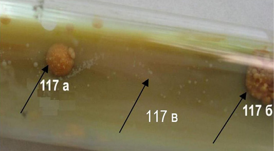
Figure 1: Colonies M. bovis of the first generation, cultivated at a temperature of 3°C (117a, 117b, 117c).
The consistency of the colonies was fragile and dry, transparent - matte, and emulsification - moderate.
By means of microscopy was established (Figure 3), that all cells of M. bovis variant 117-c of the first-generation were kept the paint of fuchsin (acid-resistant), 117-a variant - partially (in some events also had been found non-acid resistant cells), 117-b - all non-acid resistant variants (forms) of the microorganism. At the same time in the second generation of the colonies formed: only non-acid-resistant forms (117-b, c); non-acid resistant and acid-resistant (single) mycobacteria (117-a generation). In the following subcultures, beginning with the third, only non-acid-resistant mycobacteria were detected.
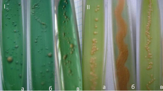
Figure 2: Culture M. bovis of dissociative forms a passage 117th, that has been formed at a temperature of 3°С: I-two weeks, II-two to four weeks, the second generation (a, b, c).
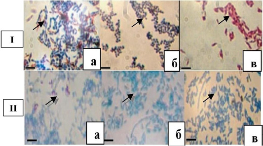
Figure 3: Dissociative forms M. bovis 117th subculture of the first (1) and the second (II) generations. Bar = 10 microns (a, b, c).
All microorganisms of the being studied subcultures, both first and second generations, had the shape of straight rod-shaped bacteria with rounded ends. The length of the microorganisms did not reveal any significant difference: short mycobacteria were dominated. This also applies to the thickness: in the vast majority, thin, sometimes thin and thick forms of mycobacteria mostly appeared in the first generation. However, mycobacteria of the first generation turned out to be longer than the traditional in the general background of short coconut like forms, which were placed by aggregations. In the second generation it is noted in the majority of single and aggregate short forms.
By studying the subcultures of the third generation, different growth rates were established on the dense nutrient medium at temperatures of 3°C and 37°C of cultivation. Almost twice as fast colonies were formed at 3°C (3-4 day) than at 37°C (5-10 days). The surface of subcultures was smooth and moist. However, they all formed a pigment: at 3°C – cream-coloured, 37°C – orange. Under the immersion of the microscope, mycobacteria from the colonies, which were obtained at 3°C and 37°C, were non-acid-resistant, short, thin, straight, with rounded edges and granular. However, in the curtain suspensions of mycobacteria, prepared from cultures that were obtained at 3°C have been revealed non-acid-resistant elementary small bodies.
Dissociative forms (117-a, b, c) cultivated and accumulated at 3°C and 37°C did not cause at guinea pigs the pathological and anatomical changes of typical tuberculosis and allergic reactions to PPD-tuberculin for mammals. The weight of the body of the experimental animals corresponded to the physiological norm. There were no ulcers in the area of introduction of mycobacteria suspension.
M. bovis 100th reinoculation in guinea pigs (control) stimulated the development of the infectious process, weight loss, allergic condition and ulcers in the inoculation zone of mycobacteria curvature. The death of animals from tuberculosis was marked on the 32nd and 42nd day of the experiment. At 10th reinoculation of mycobacteria the rate of formation of subcultures (colonies) at both temperatures of cultivation was the same – 4-7 days. Colonies of all dissociative variants are classified into S-shapes, pigmented for both 3°C and 37°C cultivation. M. bovis of dissociative forms were long and short, with the exception of the mycobacteria of the 117-a subculture of the 37°C cultivation temperature, where short, filiform forms with dark grains were established along the entire length of the cell. The thickness of mycobacteria was the same: thick and thin.
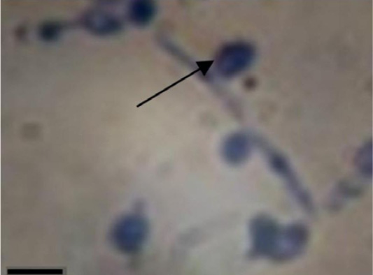
Figure 4: M. bovis of dissociative forms 117-b (10th generation), cultivated at temperature 37°C. Bar=10 microns.
However, M. bovis 117-c (at 37°C) and 117-b, c (at 3°C and 37°C) variants differed (Figure 4): thick, slightly oval rod-shaped bacteria, filiform shapes, in which dark blue grainy formations were contained. The latter, in some cases, pushed out from such cells, from which later formed typical L-shapes (ovals with different optical density of the surface).
In the form of mycobacteria – curved, straight, granular (almost equal in proportion), their ends are rounded. All cultures contained elementary small bodies (non acid-resistant), but only this was not the case for cultures that were cultivated at 37°C. L-forms of mycobacteria (protoplasts/ spheroplasts) were found in 117-b, c variants that were cultivated at 37°C and in subculture 117-c variant at 3°C.
The contamination of guinea pigs, rabbits, and chickens with mycobacteria of dissociative forms of 10th subculture was accompanied by sensitization of the macroorganism in relation to PPD-tuberculin for mammals in 50% of experimental animals, in particular guinea pigs. Some of them (for No 14) responded to the 30th and 60th day of the experiment. At the same time, none of the experimental animals showed pathological anatomical changes characteristic for tuberculosis. Guinea pigs and rabbits that are infected with the pathogenic strain M. bovis responded to tuberculin (Table 1).
The death is noted: guinea pigs for 35-48 days; rabbits 60-72 days; hens did not get sick. Only six cultures (50.0%) were isolated from bacteriological investigations of biological material from each separately tested guinea pig. Only three cultures cultivated at 3˚C, only one (16.7%) cultivated at 37°C, in cultivation simultaneously at 3°C and 37°C were two (33.3%) cultures. Morphologically, mycobacteria under the immersion system were characterized, as a rule, by grains, non-acid-resistant rod-shaped bacteria (117-a, No. No. 3-4, 117-b, No. No. 13-14), L-forms of protoplast type (117-c, No. 25), and only L-forms (117c, No. 28).
Two cultures were obtained at 3°C and 37°C simultaneously from mammals infected with mycobacteria (No. 4, 14) accumulated at 3°C and 37°C, respectively. Three cultures were obtained only at 3°C (No. No. 3, 13, 28), of which 117-a, c cultures were accumulated at 37°C, one – 117-b at 3°C. One culture (117-c) was isolated at 37°C from guinea pig (number 25) infected with mycobacteria, which was accumulated at 3°C. At the same time, as can be seen from the table, the dependency of the morphology of isolated microbial cells and the temperature of their cultivation were not detected.
The rabbits and chickens infected by M. bovis did not react to tuberculin tuberculosis with dissociative forms, and bacteriological investigations turned out as negative. Mycobacteria of the 20th subculture compared with the 10th changed the rate of reproduction (formation of colonies), especially at 37°C. At this temperature, all variants of the studied subcultures form miserable, but macroscopically visible colonies in the form of continuous growth along the line of inoculation of suspension on the second day of cultivation, while the growth of the culture of the tenth generation was marked for 4-7 days. The same subcultures were formed at the temperature 3°C, but only on the fourth day of cultivation. A week after the two temperature regimes, the subculture was formed in the form of thick continuous growth. The surface of subcultures was smooth, though, after 2-4 weeks of cultivation, individual rough colonies climbed over continuous growth.
On the 2-4 days of cultivation of dissociative forms of 20th generation at both temperatures the cream pigment was formed at 21 days – at 37°C it remained creamy, and at 3°C it acquired orange color.
In the morphology and tinctorial properties of M. bovis 20 subculture, a certain dependence on the temperature of the cultivation was observed: 1) non-acid-resistant M. bovis dissociative forms were usually generated at a low positive temperature in the dynamics of the reinoculations, as a rule, generated (with the exception of 117-a and b variants); 2) elementary bodies were formed at 37°C cultivation (except for the 117-a variant of mycobacteria, where the bodies were generated at 3°C); 3) L-forms of the protoplast type with different optical density of the surface more stably generated at 37°C with the simultaneous formation of elementary acid-resistant bodies, at 3°C against the background of a certain change in the morphology of L-forms, non-acid resistant, short and long rod-shaped bacteria (elementary small bodies are absent).
At the same time, studying the possibility of multiplying in mycobacteria of the dissociative forms of generation 25 on simple nutrient media and on the Lövenstein-Jensen medium containing 0.5% and 1% by volume of salicylic acid, it was noted that all the studied mycobacteria (117-a, b, c) formed colonies at 3°C and 37°C for 2-4 days. Pathogenic mycobacteria (maternal strain) did not show growth in the medium except for such Lövenstein-Jensen medium (without sodium salicylate), in which colonies were formed on the 14th day of incubation.
Under imersium, in smears prepared from weekly cultures obtained on agar, in the broth (film) and on Lövenstein-Jensen medium with sodium salicylate have been revealed differences in morphology and tinctorial properties of mycobacteria forms.
In the culture of the variant 117-a on the FPA at 37°C, only acid-resistant elementary small bodies and L-forms of the protoplast type were observed, in the culture on the FPB, at the same temperature, only acid and non-acid resistant grains and L-forms, while at temperatures of 3°C in the culture on the FPA registered non-acid resistant rod-shaped bacteria and acid-resistant grains, and in the FPB - non-acid resistant rod-shaped bacteria and non-acid-resistant grains.
Non-acid resistant rod-shaped bacteria and acid-resistant grains were found in 117-b subculture at FPA and FPB at temperature of 3°C, at 37°C in FPA - non-acid resistant rod-shaped bacteria, acid-resistant elementary small bodies, non acid-resistant grains and L-shapes, and only non-acid-resistant grains on FPB. Non-acid-resistant rod-shaped bacteria of different shapes and sizes and non-acid-resistant grains were found in 117-c subculture obtained at FPA at 3°C, and at 37°C there were filamentous forms and L-forms of two variants: a) large oval formations with different optical density of the surface; b) in two to three times smaller, similar in morphology to the previous formations. The latter, as shown in Figure 5, are pushed out of filiform shapes. Grain sticks and acid and non-acid-resistant grains were found on the FPB at 3°C; at 37°C only acid and non-acid-resistant grains were found.
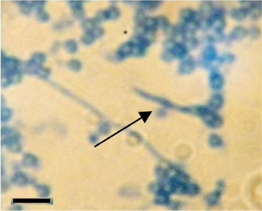
Figure 5: M. bovis 117-c variant of dissociative forms cultivated at 37°C on flesh-peptone agar (5 generation of weekly culture). Bar = 10 microns.
Consequently, in cultures on simple nutrient media at 37°C, dissociative forms were predominantly L-shaped, while at 3°C – sticks of different shapes and grains (although there were also L-forms).
Having obtained such results, we conducted repeated studies to find out the influence of the temperature of cultivation on the morphology of the dissociative forms of M. bovis, with reinoculation them to the same dense Lövenstein-Jensen nutrient medium 20 times.
The reinoculation from 3 to 23 generation confirmed the influence (Figure 6) of the temperature on the morphology of mycobacteria: in all three variants of M. bovis at 3°C the morphology remained practically stable grains, short rod-shaped bacteria were constantly generated during 20 reinoculations, whereas at 37°C sticks, grains were replaced by L-shapes and elementary small bodies.
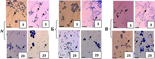
Figure 6: Morphological signs and tinctorial properties of M. bovis variants 117-a, b, c of dissociative forms, passaged through dense nutrient medium, cultivated for: left column 3°C; rights - 37°C. Bar: 10 microns.
Under such conditions, the pattern of the culture changed over time. In particular, with the advent of elementary small bodies in culture after 4-5 days of incubation, the film of continuous growth thinned, through 2-4 weeks the medium was thinned, while at 3°C the culture in the form of continuous growth along the line of inoculation of the suspension of mycobacteria intensively increased in time.
Investigating the morphogenesis of dissociative forms of mycobacteria (variant 117-c), isolated from the biomaterial of experimentally infected guinea pigs, it was established that colonies at 3ºC cultivation formed (on the 10th day of colony growth) only oval-shaped forms (Figure 7-1) did not contain grains and elementary small bodies around them. Seven days later (Figure 7-2), after repeated microscopy, spheroplasts (L-forms) acquired different optical density of the surface with clearly visible grains inside, a significant number of elementary small bodies near and around them.
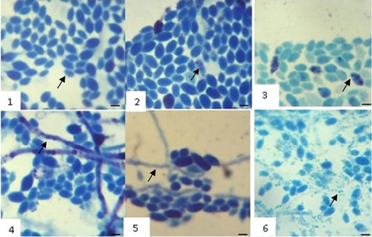
Figure 7: Initial and subsequent stages of separation of nuclear substance in M. bovis spheroplasts and formation of morphological forms: 1 - on 7; 2 - on 14; 3 - on 21; 4 - on 28; 5 - on 35; 6 – on 240 day of colony growth (117-v variant, 11 subculture isolated from guinea pig biomaterial). Bar: 10 μm.
After 21 days (Figure 7-3), the intensity of separation the nuclear material in spheroplasts (grains, elementary small bodies) significantly increased. This may indicate the persistence in the macroorganism of single mycobacteria in the form of spheroplasts that are capable of cultivating in the future on the dense nutrient medium at 3ºC with the separation of nuclear material (elementary small bodies).
After studying the morphology of the transformed M. bovis of the same colony for the next 14 days, it was revealed (Figure 7-4, 5) law-based dynamic changes that were characterized, along with the morphological forms previously given, an increase in the number of elementary small bodies, the formation of filiform variants, in which the grains (large and small) and oval-shaped formations with the same optical density of the surface that were freed from them and were clearly observed. On the 240th day of cultivation, the fluffy threads with the contents of grains (Figure 7-b), which were pushed out and transformed into L-forms (oval formations with different optical density of the surface), were established, and a sharp increase in the number of submicroscopic and microscopic, as in previous studies, non-acid-resistant elementary small bodies.
These data confirm once again that, under the condition of investigation, in the dynamics of growth of one colony, which was formed from one L-form of the spheroplastic type, the formation of spheroplasts occurs precisely from the filiform variants of M. bovis, although, in addition, in these structures, grains (elementary small bodies). At the same time, our investigations have not revealed a clear mechanism for the formation of filiform variants of M. bovis, that is, from which morphological forms they are formed. Most likely, one can only assume that from the elementary small bodies, since other formations in the investigated colony were not detected, but they, elementary small bodies (nuclear matter) were separated in spheroplasts: the threads were not observed until the appearance of elementary small bodies in the colony.
Generally, phenotypic changes in mycobacteria are closely related to the chemical composition of the cell, which, depending on the medium, varied by defining morphology, tinctorial properties, biochemical activity that contributed to adaptation to survival. At the same time, Table 2 shows that the content of total lipids in investigated mycobacteria strains was significantly different.
Their highest content was found in M. bovis of the pathogenic epizootic strain of the second subculture, however, it was lower by almost two and three times in the reference strain
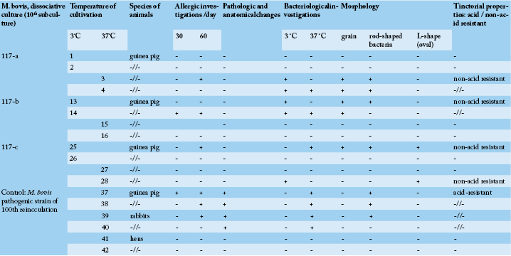
Table 1: Biological effect in experimental animals depending on infection with dissociative forms M. bovis.
Table 2: Total lipids of mycobacteria, % by weighed portion.
| Indicator | Sample | ||||||||
| 37°C | 3°C | ||||||||
| Strain of the passage | |||||||||
|
2 nd |
Vallee |
100+124/2 |
variant 117 |
||||||
| а | b | c | а | b | c | ||||
| Total lipids |
15.13±0.30 |
8.82±0.79* |
4.85±0.45*** |
2.40±0.26*** |
2.2±0.31*** |
2.6±0.32*** |
2.31±0.11*** |
2.96±0.37*** |
2.38±0.14*** |
* P≤0.05; ** P≤0.01; *** P≤0.001.
Table 3: Composition of mycobacterium lipids (thin-layer chromatography /TLC/ analysis), % of the sum.
| Fraction of total lipids | Sample | ||||||||
| 37°C | 3 °C | ||||||||
| Strain of the passage | |||||||||
|
2nd |
Vallee |
100+124/2 |
variant 117 |
||||||
| a | b | c | a | b | c | ||||
| Phospholipids |
26.18±0.25 |
23.17±0.94* |
21.69±0.43*** |
19.22±0.22*** |
20.5±0.37*** |
19.11±0.44*** |
19.2±0.58*** |
20.57±0.50*** |
20.0±0.70** |
| Diacylglicerols |
15.14±0.22 |
17.08±0.60* |
15.18±0.06 |
15.79±0.47 |
14.1±0.44 |
16.03±0.65 |
16.97±0.26** |
17.69±0.37** |
18.18±0.48** |
| Sterins |
13.56±0.31 |
17.68±0.65** |
14.78±0.25* |
17.45±0.46** |
15.81±0.51* |
16.40±0.44** |
17.34±0.59** |
16.97±0.26** |
16.37±0.51** |
| Free fatty acids |
14.2±0.25 |
16.46±0.54* |
15.77±0.52 |
14.68±0.42 |
16.67±0.39** |
16.02±0.64 |
16.97±0.26** |
15.52±0.45 |
16.36±0.50* |
| Triacylglycerols |
16.72±0.32 |
13.41±0.52** |
16.69±0.49 |
20.33±0.68** |
16.24±0.45 |
16.44±0.45 |
16.24±0.37 |
16.61±0.35 |
15.64±0.44 |
| Athers of sterols |
14.2±0.25 |
12.2±0.45* |
15.89±0.66 |
12.53±0.40* |
16.68±0.39** |
16.0±0.67 |
13.28±0.53 |
12.64±0.4* |
13.45±0.48 |
|
∑ lipids |
100 | 100 | 100 | 100 | 100 | 100 | 100 | 100 | 100 |
* P≤0.05; ** P≤0.01; *** P≤0.001.
of the Vallee and in the pathogenic mycobacterium subculture of 100-124 passages, respectively. At the same time, the dissociative forms of M. bovis in the number of total lipids, compared with pathogens, were more significantly different from the original pathogenic strain than the reference Vallee and passage 100-124 times with the highest probability. It is characteristic that, irrespective of the temperature of cultivation, the dissociative forms of M. bovis contained practically the same amount of total lipids: from 2.2% (117-b, 37°C) to 2.9% (117-b; 3°C) on the weighed portion.
Differences in the fractional composition of the lipids of strains of mycobacteria were not detected (Table 3). While in the quantitative content, probable changes were noted mainly in phospholipids, which were most pronounced in M. bovis of the second subculture of the epizootic pathogenic strain.
Also, high content of phospholipids was noted in Vallee strains mycobacteria and 100-124 subculture, in comparison with the dissociative forms of M. bovis, which contained a much smaller number of phospholipids than the pathogenic strain of mycobacteria (2 passage).
Meanwhile, diacylglycerols dominated over such 2 passages in 117-a, b, in the variants cultivated at 3°C. However, at 37°C cultivation of mycobacteria analyzed fraction did not differ from the pathogenic variants of mycobacteria (2nd passage, Vallee).
The probable difference from the source of the pathogenic strain of such fractions was also noted: sterols (more in all investigated strains); free fatty acids (more in mycobacteria of the Vallee strain, dissociative forms of 117-b (37°C) and 117-a, c (3°C); triacylglycerols (more in mycobacteria Vallee and 117а (37°C); athers of sterols (more in the Vallee strain, but probably less than those in the 117-a, b (3°C). Almost identical profiles of free fatty acids with the number of carbon atoms from C11 to C27 have been isolated (Table 4). The basic fatty acids are established: myristic, palmitic, palmitoleic.
However, in the pathogenic strains, undecanoic acid was not isolated (C11:0), and the re-inoculation of the epizootic strain 100-124 times was accompanied by the decrease to the non-diagnostic level of hexacosanic (C26:0) and heptacosanic (C27:0) free fatty acids, which were also not detected in dissociative forms of mycobacteria, cultivated for both 3°C and 37°C. Only traces of arachidonic acid (C20:4) were isolated merely in M. bovis of the second subculture of the epizootic pathogenic strain.
Trustworthy differences in the quantitative composition of free fatty acids were observed in both pathogenic strains of mycobacteria and their dissociative variants. The amount of unsaturated fatty acids was lower in the pathogenic strain M. bovis, in comparison with dissociative forms of mycobacteria, cultivated in two temperature regimes, in the interval from 1.2 to 1.8 times. However, at
Table 4: Free fatty acids of mycobacteria, % of the amount.
| Free fatty acids | Code | Sample | |||||||||
| 37°C | 3°C | ||||||||||
| Strain of the passage | |||||||||||
|
2nd |
Vallee |
100+124/2 |
variant 117 |
||||||||
| a | b | c | a | b | c | ||||||
| 1 | 2 | 3 | 4 | 5 | 6 | 7 | 8 | 9 | 10 | 11 | |
| Undecanoic |
С11:0 |
- | - | - | traces |
0.42±0.08 |
0,2±0.06 |
0.32±0.27 |
0.4±0.05 |
0.28±0.06 |
|
| Lauric |
С12:0 |
0.01±0.004 |
0.41±0.15 |
0.07±0.02 |
traces | traces | traces |
0.06±0.04 |
traces |
0.11±0.32 |
|
| Tridecanoic |
С13:0 |
0.07±0.01 |
0.25±0.12 |
0.38±0.15 |
0.04±0.03 |
0.78±0.09*** |
0.29±0.07* |
0.16±0.06 |
0.52±0.07** |
0.28±0.61 |
|
| Myristic |
С14:0 |
0.16±0.02 |
0.41±0.10 |
0.31±0.13 |
0.76±0.09** |
1.3±0.10*** |
0.44±0.05** |
0.6±0.30 |
1.6±0.60 |
0.53±0.21 |
|
| Pentadecanoic |
С15:0 |
0.16±0.04 |
0.36±0.13 |
0.63±0.12* |
0.19±0.04 |
1.3±0.10*** |
0.58±0.08** |
0.57±0.29 |
0.48±0.05** |
0.5±0.30 |
|
| Palmitic |
С16:0 |
25.52±0.45 |
18.87±0.98** |
27.67±0.68 |
25.22±0.39 |
39.81±0.95*** |
45.59±0.58*** |
33.76±0.89*** |
26.12±0.53 |
23.95±0.06** |
|
| Margarine |
С17:0 |
traces |
1.27±0.20 |
0.98±0.11 |
11.33±0.78 |
0.26±0.07 |
0.29±0.01 |
0.82±0.46 |
4.27±0.61 |
traces | |
| Stearic |
С18:0 |
11.28±0.26 |
11.75±0.59 |
15.42±0.64** |
25.98±0.57*** |
18.27±0.75*** |
31.39±0.32*** |
16.58±0.30*** |
11.8±0.93 |
14.6±0.21*** |
|
| Nonadecanoic |
С19:0 |
0.73±0.05 |
2.31±0.50* |
2.97±0.58* |
0.02±0.01*** |
traces |
0.19±0.01*** |
0.69±0.31 |
3.56±0.27*** |
1.28±0.30 |
|
|
Аrаchic |
С20:0 |
4.38±0.27 |
2.68±0.37* |
0.13±0.06*** |
1.21±0.53** |
1.74±0.17** |
2.21±0.19** |
13.39±0.24*** |
0.45±0.42** |
11.48±0.96** |
|
| Heneicosanic |
С21:0 |
6.16±0.23 |
5.13±0.43 |
0.78±0.17*** |
0.76±0.48** |
0.52±0.07*** |
0.72±0.07*** |
0.69±0.31*** |
0.59±0.33*** |
0.8±0.36*** |
|
| Behenic |
С22:0 |
6.18±0.05 |
5.21±0.49 |
2.86±0.52** |
2.58±0.56** |
2.62±0.08*** |
1.3±0.09*** |
1.26±0.51*** |
4.08±0.59* |
1.95±0.14*** |
|
| Tricosanic |
С23:0 |
traces |
3.31±0.57 |
0.17±0.07 |
1.13±0.64 |
0.04±0.01 |
0.04±0.01 |
0.03±0.04 |
0.89±0.24 |
0.57±0.32 |
|
|
Теtracosanic |
С24:0 |
3.6±0.20 |
2.9±0.81 |
0.06±0.02*** |
0.67±0.33** |
0.17±0.01*** |
0.14±0.03*** |
0.86±0.46** |
traces |
0.28±0.24*** |
|
| Pentacosanoic |
С25:0 |
15.84±0.29 |
12.72±1.05* |
5.27±0.70*** |
0.38±0.35*** |
0.09±0.02*** |
0.04±0.01*** |
0.57±0.29*** |
0.9±0.24*** |
0.43±0.06*** |
|
| Hexacosanic |
С26:0 |
1.12±0.05 |
1.46±0.46 |
- | - | - | - | - | - | - | |
| Heptacosanic |
С27:0 |
0.09±0.03 |
traces | - | - | - | - | - | - | - | |
|
∑ saturated acids |
75.3±0.23 |
69.04±1.40* |
57.7±0.80*** |
70.27±0.48*** |
67.32±0.32*** |
83.42±0.88*** |
70.36±1.08* |
55.66±0.48*** |
57.04±1.21*** |
||
| Palmitoleic |
С16:1 |
0.87±0.04 |
0.99±0.32 |
1.11±0.38 |
0.02±0.01*** |
0.22±0.09** |
0.12±0.01*** |
0.51±0.31 |
1.14±0.50 |
1.2±0.14 |
|
|
Оleic |
С18:1 |
23.29±0.56 |
27.18±1.43 |
39.81±0.72*** |
6.15±0.44*** |
22.2±0.62 |
13.2±0.56*** |
28.49±1.27* |
39.63±0.45*** |
40.17±0.57*** |
|
|
Linoleic + linolenic |
С18:2+ С18:3 |
0.54±0.08 |
2.79±0.85 |
1.38±0.19* |
23.56± 0.48*** |
10.26±0.59*** |
3.26±0.36** |
0.64±0.31 |
3.57±0.27*** |
1.59±0.28* |
|
| Arachidonic |
С20:4 |
traces | - | - | - | - | - | - | - | - | |
|
∑ unsaturated acids |
24.7±0.43 |
30.96±1.39* |
42.3±0.82*** |
29.73± 0.61* |
32.68± 0.53*** |
16.58± 0.50*** |
29.64±1.17* |
44.34± 1.11*** |
42.96± 1.14*** |
||
* Р≤0.05; ** Р≤0.01; *** Р≤0.001.
mycobacterium in the variant 117-c cultivated at 37°C, this indicator was 0.7 times smaller. Such changes in the content of unsaturated fatty acids were conditioned of the synthesis of oleic acid (C18:1); and in the variant 117-a at 37°C – the synthesis of the amount of linoleic and linolenic acids.
The obtained data testify to the identity of the qualitative composition of free fatty acids in the pathogenic and dissociative forms of M. bovis, which confirms the origin of dissociate from the maternal strain. At the same time, due to the constant adaptability to environmental factors, the content of unsaturated fatty acids was quite high from the multiple passages of M. bovis strains (100-124) and the referential strain of Vallee M. bovis.
DISCUSSION
After the first report by Klienberger-Nobel in 1935 about the ability of Streptobacillus moniliformis to change morphologically (L-forms), information on this phenomenon appeared in other bacteria (Klienberger, 1935; Dienes, 1939), including mycobacteria. In this type of microorganism, both under the influence of inducing factors and in macroorganism (although here the internal environment affects the adaptive capacity of mycobacteria), there is the conversion in the L-form (cells with a cell wall deficiency, and morphologically different from typical acid-resistant rod-shaped bacteria). At the same time, a number of researchers (Beran et al., 2012; Markova et al., 2012) believe that mycobacteria can be converted into L-forms naturally, without the influence of inducing factors, that is, this process is genetically embedded in the microbial cell and is aimed at survival.
In this investigation the mechanisms of changing the morphology of the microbial cell at low positive temperatures (3°C) are demonstrated in conditions of numerous reinoculations through the dense nutrient medium on the background of qualitative and quantitative lipids changes, which can be used to determine the role of L-forms in long-term survival in the environment. The investigations with this methodological approach have not been found by us that emphasizes their peculiarity and significance, especially when dissociation from the maternal pathogenic strain of nonpathogenic cells is capable of demonstrating other biological properties, but retaining the biological cycle of development, which has been researched by other researchers under the influence of inducing factors and without them (Domingue and Woody, 1997).
However, if there is enough information on the influence of artificial induction, limited information is available on such the dense artificial nutritional medium (Kochemasova, 1980). Usually this characterizes mycobacteria, in particular M. bovis, as a fairly labile microorganism that is capable of transforming, reorganizing the metabolism depending on the environment, keeping the viability uncertain for a long time. Up to 116th reinoculation mycobacteria on artificial dense nutrient medium with pH 7.1 has typical biological properties for M. bovis.
Meanwhile, many month storage of culture at the margin of visibility in the form of “haze”, “costing” in the conditions of low positive temperatures, not inherent to the life of mycobacteria without reinoculation to the support medium, influenced the metabolism of the microbial cell and determined the mechanism of survival under such conditions. It was hypothetically assumed that multiple reinoculation and especially storage of mycobacteria cultures in the conditions that were unsuitable for optimal metabolism of mycobacteria led to the dissociation of certain microbial cells of the pathogen of tuberculosis, which began to propagate at temperatures 2-3°C for 20-months of cultivation. This confirms the certain reorganization of the genetic code in individual cells of the studied mycobacteria.
If, in the absence of inducing factors in the population of mycobacteria, as reported by the authors (Udou et al., 1982; Markova et al., 2012), the gradual appearance of L-shaping cells is observed: transitional non-acid resistant rod-shaped bacteria, filamentous variants, L-forms of the protoplast (spheroplastic) type, grains (elementary small bodies), then in our research dissociation phenomena were accompanied by a one-time appearance in individual cells of the ability to transform into non-pathogenic mycobacteria, while the maternal population of cells continued to generate pathogenic micrograms.
A small amount of dissociation of mycobacteria (23 passages through artificial dense nutrient media) was accompanied by the influence of the temperature of cultivation on their morphological characteristics: stable non-acid-resistant short and long non-shaped bacteria were stably generated at 3°C, and the morphology of the L-forms of the protoplast type was slightly changed; at 37°C elementary small bodies and L-shapes. At the same time, our research confirms the number of reports of previous years (Dines, 1939; Domingue, 2010; Markova et al., 2012), about that, the mycobacteria, under the influence of inducing factors, are transformed, defining a certain biological cycle of development, and in particular, in the beginning arise the adaptive forms non-acid resistant non-shaped bacteria, thread-like variants, from which (the latter) oval-shaped forms with ripening grains (elementary small bodies) are pushed out, oval-shaped variants, that is, spheroplated L-shapes, collapsing to liberate elementary small bodies. Such bodies, according to the author’s information (Markova et al., 2012), are the final stage of the biological cycle of the development of mycobacteria. They (the bodies) can give beginning to the rod-shaped variants of mycobacteria.
An illustrative phenomenon was established by us at cultivation of dissociants on the simple nutrient medium (the variant of culture 117-c), and the variant of mycobacteria 117-b cultivated on an elective dense medium.
Usually more objective and representative are the investigations of the biological cycle of the development of mycobacteria from one cell, from the stage of spheroplasts isolated from biological material of guinea pig and studied (cultivated at 3˚C) in the dynamics of growth of one colony. The established cycle sufficiently demonstratively testifies the formation of spheroplasts from filiform variants. Spheroplasts separate the large amount of nuclear material in the form of elementary small bodies, which are then pushed out of the vesicular form as multiplying organisms.
These results fully confirmed the information (Domingue and Woody, 1997), who believes that one large vesicular maternal mature form may develop into the large number of elementary small bodies that are then pushed out of the maternal form. At the same time, we did not find the mechanism of formation of filamentous forms, that is, of which elements they formed in the dynamics of colony growth. Meanwhile, the authors’ report (Udou et al., 1982) shows that observing in the electron microscope the formation of Mycobacterium smegmatis spheroplasts and the morphological aspects of their reversion in bacillary forms was found two obvious ways of reversing. The first way of these was initiated by the budding of spheroplasts: the buds gradually elongated, becoming the micellar forms, which manifested branching, the appearance of partitions and fragmentation. The second way came from the intracellular formation of tiny little cells, possibly elementary small bodies, and their release from spheroplasts.
These changes, as have been evidenced by our examinations, are given in this work, accompanied by adjustment of the microbial cell metabolism. This led to the possibility of multiplication and accumulation on simple nutrient media and with the content of sodium salicylic acid. Although it is known that pathogenic mycobacteria are not cultivated in chemically simple media (Ramakrishnan et al., 1972; Narvaiz de Kantor et al., 1998). Such variants of mycobacteria that are able to survive in an environment that is not inherent to them (in conditions of low positive temperatures), with gaining, increasing the metabolism by properties, are closer to atypical mycobacteria, which, like the experimental variants of mycobacterium, which are investigated, do not cause death of guinea pigs, rabbits and chickens, however, can cause the condition of the infection and stimulate a latent infectious process in animals, in particular bovine animals, as reported by Tkatschenko and Schewziw, 1994, in which an allergic reaction can / cannot be shown and tuberculin, which was found in these research when infected laboratory animals in experiments 25% did not respond to allergen for mammals.
The rabbits and chickens did not show any sensitivity at all to the diagnostic preparation. At the same time, the long-term (3 months) persistence of altered different morphological forms of mycobacteria in the body of guinea pigs can testify to their stimulation of antituberculous immunity. Usually, to prove this, the following experiments with the appropriate methodological approach are required.
Of course, the morphology and pathogenic ability of mycobacteria is connected with the chemical composition, in particular, with lipids. It is possible, it can be argued that only single microbial cells over a 20-month stay in the low positive temperature changed the metabolism, sharply reduced the content of total lipids, and free fatty acids displaced by the number of carbon atoms towards those with low content and synthesis to the diagnostic level to this uncertified undecanoic free fatty acid. On this background, the ability of mycobacteria to synthesize substances decreased / or they are not delayed in a friable cell membrane, which determine acid resistance, that is, chemical agents that in virulent mycobacteria (the parent strain) delay the fuchsin in their membrane. Usually low lipid content in the cell wall of the virulent strain of mycobacteria (2 subcultures) increases the possibility of penetration into the microorganism of damaging substances, but at the same time, increases the ability of metabolic processes, which ensure fast reproduction of dissociative forms of mycobacteria for the second to third day of cultivation.
Possibly this may change the activity of individual synthetase systems, aimed at changing the biological properties of the microbial cell, its ability to rapidly multiply, with the preservation in time of passages through artificial dense nutrient medium, high virulence of the microbial cell. Just the low content of lipids and the displacement of activity of the corresponding genes of the microorganism determined the dissociation of pathogenic (avirulent) microbial individual cells from the pathogenic parent strain in extreme conditions of stay. In such mycobacteria there is the decrease in the activity of individual genes, stimulating the certain biological activity with the corresponding properties and simultaneous activation of “dormant”. Last activating determines certain other properties. This phenomenon is clearly demonstrated in mycobacteria of our research, which is evidence of the general biological property.
Downing et al., 2005; Tufariello, 2004; Yeremeev et al., 2003 conform that the “resuscitation-promoting factor” or the rpf genes play an important role in alleviating M. tuberculosis, assuming that they are essential for returning from sleep state.
The confirmation of these data also indicates the following. Grown in vitro in conditions approximate to in vivo, M. tuberculosis is adapted by special sets of partially overlapping genes (Sherman et al., 2001; Voskuil et al., 2003; Yuan et al., 1996). Increased expression of genes such as HspX (so-called α crystalline, Rv, 2013 c, etc.) during the stationary phase of growth (Yuan et al., 1996) is critical to the survival of the microorganism (Yuan et al., 1998).
In concordance with these observations in vitro, it has been shown that transcription of a number of genes was regulated with the decrease, while others are regulated in infected animals strictly with an increase after initiation of a strong immune response (Shi et al., 2003). Together with the excess of regulatory proteins in the M. tuberculosis genome (Cole et al., 1998), this shows the importance of the ability of the pathogen to adapt during the infection to different environmental conditions. This regulatory flexibility may underline its ability to switch between acute progressive disease and long-term latent infection.
At the same time, acids with 26-27 carbon atoms are not identified. The shift in metabolism in the cell causes the processes of conversion of mycobacteria to other forms, which determines the biological cycle of the development of the pathogens of tuberculosis.
Instead, Taneja et al., 1979 reported that in the investigation of the influence of the cultivation temperature on the lipid content of Mycobacterium smegmatis ATCC 607, the total content of lipids and diacylglycerols did not change depending on the temperature of cultivation (27°C or 37°C). However, the content of phospholipids, triacylglycerols and monoacylglycerols, unsaturated fatty acids from total phospholipids significantly increased at lower temperatures.
The similarity of chromatograms in different strains of the same species and their differences in different species is also indicated by the researchers Thoen et al., 1971a; Larsson and Mards, 1976; Tisdall et al., 1979; Nandedkar, 1982, 1983.
In addition, Thoen et al., 1971b confirm that pathogenic and nonpathogenic strains, both freshly allocated and those that persisted for a long time (2 years), were similar in fatty acid profiles, but different in C14:0, C16:1, C18:0, C18:1 and branched acids among strains of one serotype.
Among other things, some researchers report that changes in the amount of unsaturated to saturated fatty acids are noted or not observed in the dynamics of the reinoculation and storage, which may indicate the preservation or loss of pathogenicity of the microorganism. So, in Mycobacterium phlei, reports Suutari and Laakso, 1993, the unsaturation of fatty acids was increased at the decrease in temperature (from 35°C to 26-20°C).
Our investigations confirm, that the significant amount of reinoculation through an elective dense medium at the low positive cultivation temperature (2-3°C) leads to the increase in the content of unsaturated fatty acids, on the background of significant loss of pathogenicity of mycobacteria that were investigated.
Of course, this is connected with the adaptation of mycobacteria to stressful events occurring at constant multiple reinoculations and low positive temperatures. In the maternal pathogenic strain M. bovis of the second generation, after isolation from biological material, the amount of saturated acids in 1.1 -1.3 times higher than that of the passaged strain Vallee M. bovis and strain BCG M. bovis, which also repeatedly passaged, which is arguably confirmed our view that passages (adaptation) affect the metabolism of the microbial cell, determining the biological cycle of development with an appropriate level of saturated / unsaturated free fatty acids in those or other morphological forms of the transformation stages of the microbial cell.
CONCLUSIONS
1. In the conditions of low positive temperatures (2-3°C) from M. bovis of the epizootic strain are split off apatogenous cells, which differ in morphology, cultural and tinctorial properties.
2. Dissociation of M. bovis is accompanied by the change in the metabolic processes of the microbial cell, which leads to a wide range of survival in the environment, and in particular the synthesis of nutrients from simple media on which the modified morphological forms of M. bovis are actively propagated.
3. Dissociation forms are transformed into elective nutrient media from non-acid resistant rod-shaped bacteria, grains in the filiform, L-shaped spheroplastic (protoplast) type and elementary small bodies: at 37°C – L-shaped and elementary small bodies; at 3°C – grains and rod-shaped bacteria, indicating the stages of the biological cycle of development.
4. Adaptation of the dissociative forms of M. bovis to extreme conditions is accompanied by dynamic changes in total lipids (reduction) of the quantitative content of fractions of total lipids and an increase in the level of unsaturated free fatty acids.
AUTHORS CONTRIBUTION
Аll authors carried out the diagnosis and photo the samples, then collection the references and wrote the manuscript.
Conflict of interest
There is no conflict of interest.
REFERENCES


