Advances in Animal and Veterinary Sciences
Research Article
Adv. Anim. Vet. Sci. 8(2): 208-212
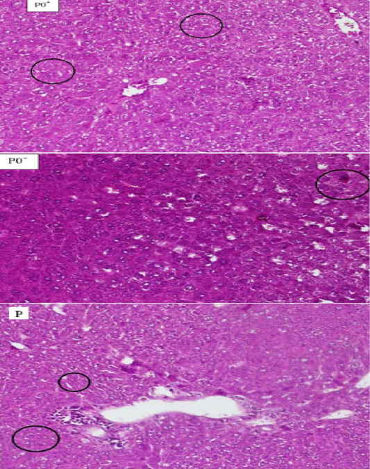
Figure 1
The comparison between hepatocyte Histopathology of mice (Mus Musculus) which experienced necrosis on the blood smears P0+, P0- and P. The circle shows the cell that experienced necrosis. The supply used Hematoxylin Eosin (HE) 200x magnification.
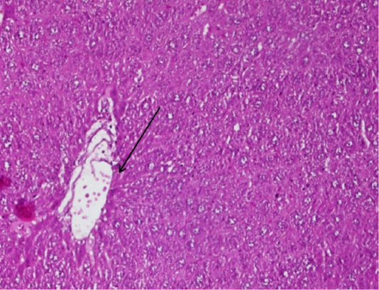
Figure 2
The arrow shows the figure of fibrosis tissue in the blood smear P of the mice (Mus Musculus) hepatic, the supply used Hematoxylin Eosin (HE) 200x magnification.


