Advances in Animal and Veterinary Sciences
Research Article
Adv. Anim. Vet. Sci. 7(s2): 183-190

Figure 1
HRTEM of chitosan nanoparticle showed nanosphere shape, no aggregation and size 26.15nm with Mag. 8000× to 16800× and 30000× to 63000× (Central lab. in NRC).
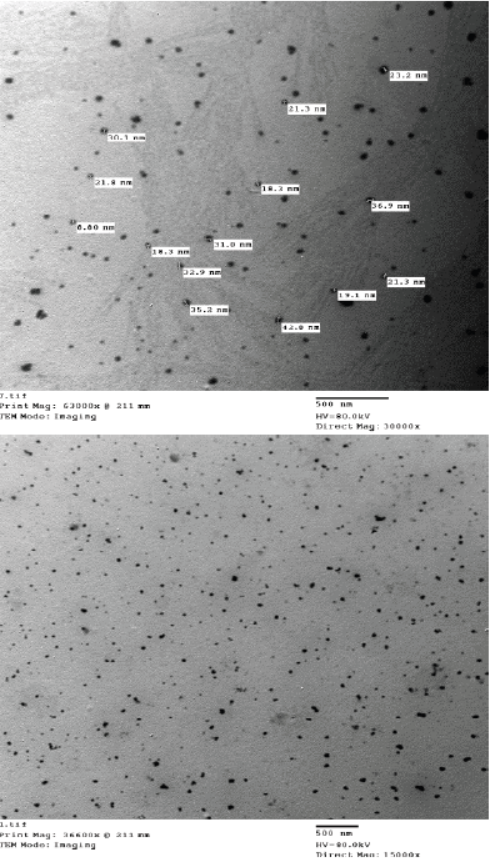
Figure 2
HRTEM of chitosan-propolis nanocomposite showed nanosphere shape, no aggregation and size 29.41 nm with Mag. 30000× to 63000× and 15000× to 36600× (Central lab. in NRC).
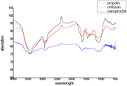
Figure 3
FTIR of propolis, chitosan and propolis-chitosan nanocomposite (Central lab. in NRC).

Figure 4
Zeta potential chitosan (A) and chitosan-propolis nanocomposite (B) (Central lab. in NRC).
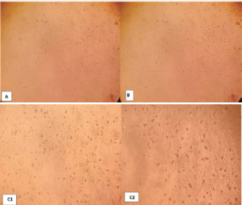
Photo 1
Negative control Vero cells 72 hr. post inoculation (A), chitosan nanoparticle no effect to cells (B) and chitosan-propolis nanocomposite (C) after 24 hrs(1) and 72hrs(2).


