Advances in Animal and Veterinary Sciences
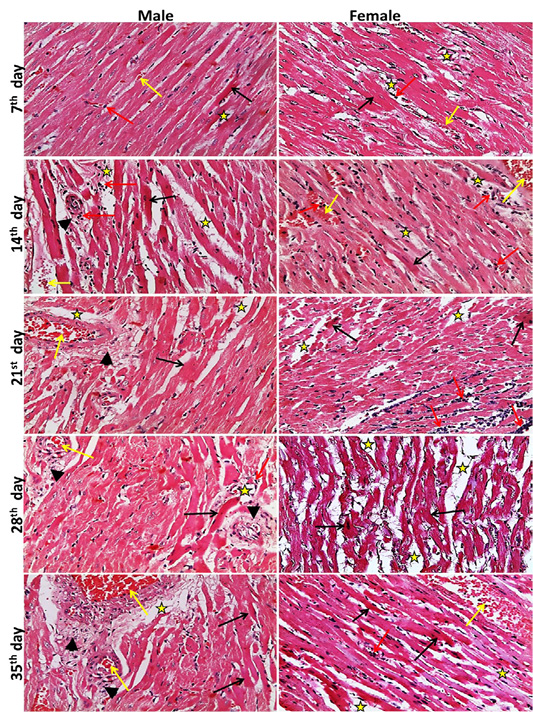
Representative photomicrographs of heart sections of male and female rats at 7th, 14th, 21st, 28th, 35th days (H&E stain X20) {Interstitial edema (stars), congestion (yellow arrows), inflammatory cell infiltration (red arrows), necrotic muscles (black arrows), perivascular fibrosis s (head arrows).
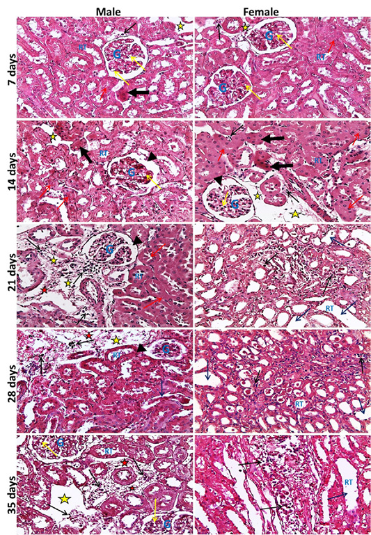
Representative photomicrographs of kidney sections of male and female rats at 7th, 14th, 21st, 28th, 35th days (H&E stain X20) {glomeruli (G), renal tubules (RT), widening of Bowmans space (head arrows), vacuolar degeneration of glomerular cells (yellow arrows), necrotic renal tubules (thick black arrows), cloudy swelling of renal tubules (red arrows), dilated renal tubules (blue arrows), inflammatory cell infiltration (black thin arrows), edema of interstitial tissue (yellow stars), fibrosis (red stars).
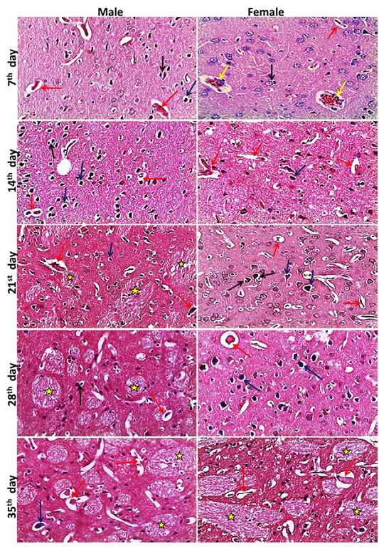
Representative photomicrographs of brain sections of male and female rats at 7th, 14th, 21st, 28th, 35th days (H&E stain X20) {Perivascular cuffing (yellow arrows), neuronophagia (red arrows), perineural satellitosis (black arrows), necrotic neurons with perineural edema (blue arrows), demyelination (stars).


