Advances in Animal and Veterinary Sciences
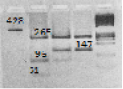
Electrophoretogram of DNA-typing of the bGH-AluI polymorphism (Skinkytė et al., 2005); Track 1 – amplificate 428 of the nucleotide pairs in a fragment of the bGH-AluI gene; track 2 – fragments of restriction 265, 96, 51 p.n. (16. p.n. are not visualized) corresponding to the bGH-AluILL genotype; track 3–fragments of restriction 265, 147, 96, 51 p.n. corresponding to the bGH-AluILV genotype; track 4–fragments of restriction 265 and 147 p.n. corresponding to the bGH-AluIVV genotype; and track 5– molecular masses marker O’Range RulerTM DNA Ladder, Fermentas, Lithuania. Electrophoresis was performed in 2% agarose gel (SeaKem LE Agarose, Lonza, USA).
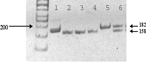
An electrophoretogram of DNA typing of the bGHR-SspI polymorphism (Zhang 1993); Track 1 – PCR product of the 182 p.n. fragment of the bGHR-SspI gene; tracks 2, 3, and 4 – a fragment of the 158 p.n. restriction corresponding to the bGHR-SspIFF genotype; track 5 – a fragment of the 182 p.n. restriction corresponding to the bGHR-SspIYY genotype; and track 6 – fragments of the 182 and 158 p.n. restriction corresponding to the bGHR-SspIFY genotype. The fragment of 24 p.n. is not visualized. The authors used molecular mass marker O’Range Ruler TM 50 bp DNA Ladder, Fermentas, Lithuania. The positions of specific bands on the gel are shown by arrows. Electrophoresis was performed in 2% agarose gel (SeaKem LE Agarose, Lonza, USA).
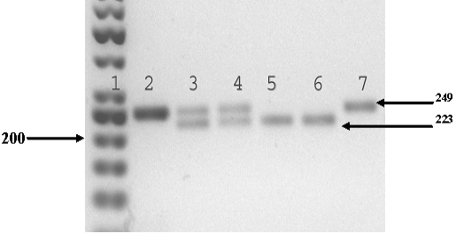
Electrophoretogram of DNA typing of the bIGF-1-SnaBI polymorphism (Kenny et al., 2011); Track 1 – molecular mass marker O’Range Ruler TM 50 bp DNA Ladder, Fermentas, Lithuania; track 2 – PCR product of the 249 p.n. fragment of the bIGF-1-SnaBI gene; tracks 3 and 4 – fragments of the 249 and 223 p.n. restriction corresponding to the bIGF-1-SnaBIAV genotype; tracks 5 and 6 – a fragment of the 223 p.n. restriction corresponding to the bIGF-1-SnaBIAA genotype; and track 7 – a fragment of the 249 p.n. restriction corresponding to the bIGF-1-SnaBIVV genotype. The fragment of 26 p.n. is not visualized. The positions of specific bands on the gel are shown by arrows. Electrophoresis was performed in 2 % agarose gel (SeaKem LE Agarose, Lonza, USA).


