Advances in Animal and Veterinary Sciences
Research Article

Hepatosis in High-Yielding Cows of the Holstein Breed
Ivan Isaevich Kalyuzhny*, Ivan Sergeevich Stepanov, Alena Alexandrovna Shimanova, Daria Sergeevna Markova, Meruert Birzhanovna Kenzhegaliyeva
Saratov State Agrarian University
Abstract | The results of the study identified the connection of hepatosis in Holstein cattle in the farms of the Saratov region and problems of feeding quality. The author has found that 38% of cows demonstrated biochemical changes in the blood serum, which is a manifestation of fatty liver dystrophy. Low level of cholesterol, sugar, vitamin A, and high content of bilirubin are the signs of this pathology.
Keywords | Holstein cattle, Metabolism, Functional liver failure, Hepatosis
Received | June 12, 2019; Accepted | August 30, 2019; Published | October 15, 2019
*Correspondence | Ivan Isaevich Kalyuzhny, Saratov State Agrarian University; Email: kalyuzhnyiii@sgau.ru
Citation | Kalyuzhny II, Stepanov IS, Shimanova AA, Markova DS, Kenzhegaliyeva MB (2019). Hepatosis in high-yielding cows of the Holstein breed. Adv. Anim. Vet. Sci. 7(s1): 9-14.
DOI | http://dx.doi.org/10.17582/journal.aavs/2019/7.s1.9.14
ISSN (Online) | 2307-8316; ISSN (Print) | 2309-3331
Copyright © 2019 Lukkananukool et al. This is an open access article distributed under the Creative Commons Attribution License, which permits unrestricted use, distribution, and reproduction in any medium, provided the original work is properly cited.
INTRODUCTION
The problem of hepatosis in high-yielding cows is one of the most acute in modern livestock breeding in the Russian Federation (Durand et al., 1998; Gruttadauria et al., 2005). The prevalence of this pathology can reach 30-40% of the total number of cattle (Doffoel-Hantz et al., 2005; Kaido and Uemoto, 2010; Kim and Sherker, 2004).
The risk of hepatosis in a herd of high-yielding cows is largely related to metabolic disorders associated with modern milk production technology (Doffoel-Hantz et al., 2005). With a high level of lactation, the maximum physiological load is taken by the liver, which has direct and indirect participation in all types of metabolism. Functional changes in its work lead to disorders, both in individual organs and in the body as a whole (Tedesco et al., 2004).
In Russia, the study of liver diseases in high-yielding cattle, in particular, hepatosis, was conducted by Zharov (1978, 1979), Postnikov (1977, 1988), Kondrakhin (1991), Baymatov (1990), Volkova (2002) among others. According to the general conclusion, manifestations of hepatosis are not specific, therefore, only a set of studies allows solving the issues of etiology and nosological affiliation of this form of liver damage (Kaido and Uemoto, 2010; Zhu et al., 2010; Guo et al., 2014; Mazzaro et al., 2003).
Taking into account certain regional peculiarities of hepatosis development in Holstein cows, which received a selection priority in the territory of the Lower Volga region, the Department of “Animal Diseases and Veterinary Sanitary Expertise” of Saratov State Agrarian University named after N.I. Vavilov carries out scientific monitoring aimed at solving the problems of combating this pathology in dairy enterprises of this region of the Russian Federation.
The set of research tasks includes the study of clinical-physiological and clinical-biochemical features of hepatosis prevalent in cows of the Holstein breed in the farms of the Saratov region; the study of the active etiological factors of hepatosis in productive cows in the region, which determine the stationarity of the dairy farming dysfunction in this pathology; clarification of the nature of additional recreational measures.
MATERIALS AND METHODS
The work was carried out in the production conditions of the breeding plant “Meliorator” CJSC in Marks district, Saratov region. Cows of the Holstein breed, at the age of 3-5 years were the objectives of the study. The research was carried out in winter, in the farm with a stall and outdoor cattle breeding.
To determine the extent of the spread of hepatosis and its causes, 1217 cows were under the dispensary control.
The methodological basis for the diagnosis of the pathology was a laboratory and biochemical analysis of the blood of a milking herd. The list of biochemical indices of peripheral blood of animals included a complex adopted to detect disorders of the hepatobiliary system in lactating cows: carotene, total protein, albumin, globulin, cholesterol, glucose, bilirubin and its fractions, liver enzymes–AST (aspartame aminotransferase) and ALT (alanine aminotransferase), alkaline phosphatase (ALP), reserve alkalinity, gamma-glutamyltransferase (GGT), vitamin A, and calcium.
Blood tests were carried out in the scientific laboratory of the Department of “Animal Diseases and Veterinary Sanitary Expertise” with the biochemical analyzer “Stat Fax 3300”, using standard reagent kits of “Diakon DS” CJSC. To check the correctness and accuracy of biochemical indices determination in the blood serum of animals, the control serum was used for biochemical studies according to Specifications 9398-022-09807247-2009, “HOSPITEX DIAGNOSTICS” LLC.
Hematological blood tests were carried out with the hematology analyzer MicroCC-20Vet, HTI (USA).
To determine the nature of the etiological factors influencing the hepatosis development, the quality of feeds and their effect on the indices of rumen digestion of lactating cows with metabolic disorders were studied. The feeds were examined in the biochemical laboratory of the State Station of the Agrochemical Service “Saratovskaya”. Laboratory studies of the rumen content were carried out according to the existing guidelines developed at the Department of “Therapy” of Saratov State Agrarian University named after N.I. Vavilov.
Statistical analysis of materials was carried out within the framework of the “Excel” program.
RESULTS AND DISCUSSION
The problem of hepatosis in cattle in the breeding plant “Meliorator” CJSC was determined at slaughter of the rejected cows, due to their loss of milk productivity. During veterinary and sanitary examination of internal organs, fatty degeneration of the liver (fatty dystrophy) was revealed, corresponding to the pathoanatomical picture of hepatosis: an increase of the organ, flabbiness, and clay color. In this case, there was generalized obesity in the subcutaneous tissue, in the pleural and peritoneal cavities, heart and kidneys. The liver with obesity had increased size, yellow color, rounded edges (Figure 1, 2).
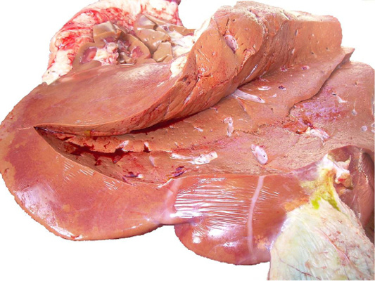
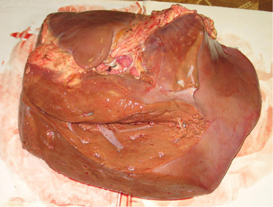
Figure 2: Fatty degeneration of the liver with chronic hepatosis in a cow rejected due to the loss of milk production.
During the examination of a dysfunctional herd, a significant part of the livestock had signs of metabolic disorders: obesity in 12% of cows, dystrophy in 9%. The general pathology in cows was recorded: osteodystrophy- resorption of the last caudal vertebrae, deformation of the hoof horn; hypotension of the forestomach; heart failure; retardation (in 17% of animals). Conjunctivitis, dermatitis, edema of the udder after calving, and necrobacteriosis were determined in the surveyed herd.
It was found out that by the end of the winter stall period, liver diseases occupied a significant place in the structure of non-contagious pathology. At the same time, an increase in the boundaries of this organ (widening of the zone of hepatic dulling) was in 52% of cows, soreness (increased pain sensitivity) during palpation in 48% of animals. Both of these symptoms occurred in 10% of cows. Hepatosis had a chronic form and latent course of the disease.
In cows suspected of the disease oppression, decreased appetite, body temperature within normal limits was determined. The color of the mucous membranes did not change (the yellowness of the mucous membranes was absent), the dermal cover was matted and ruffled. In addition, cows with signs of fatty liver dystrophy had signs of general pathology: weight loss, diarrhea, mycosis dermatitis, mastitis, endometritis, laminitis, a long service period (more than 120 days), low viability of offspring. At the same time, the highest incidence of hepatosis was in cows during 3-5 lactations (44%).
During the laboratory and clinical examination of lactating cows it was found out that a significant part of the animals (up to 42.3%) had biochemical changes in the blood characteristic for the disturbance of metabolic processes in the animals (Table 1). carotene concentration reduced in10 times (in comparison with the norm) 0.063- 0.084 mg% (in 71.7% of cows); cholesterol concentration increased by 12% - up to 6.45-6.87 mmol/l (in 65% of cows); AST activity increased up to 69.1 u/l, ALT level was normal–12.0-19.9 u/l; glucose level in all cows decreased to 1.7-1.9 mol/l; bilirubin concentration increased to 19.3-20.3 μmol/l; level of alkaline phosphatase increased in 2-3 times - 266-451 u/l (in 57% of cows). In addition, the level of total protein decreased to 54.14-70.33 g/l in 94.7% of cows. These indices are characteristic of the total protein deficiency. In 5.3% of animals, this indicator increased to 98.34 g/l.
The listed phenomena- dysproteinemia, low cholesterol, sugar, vitamin A, high bilirubin, formed a complex of signs of functional liver failure. Pigmental metabolism disorder and a decrease in the level of acid-base metabolism indicates the acidotic state of high-yielding cows. The obvious hypoglycemia, that increased to 1.7 ± 0.13 mmol/l by the end of the stall period, the decrease in protein and synthesizing liver function, as well as the simultaneous increase in cholesterol level to 6.53 ± 0.63 mmol/l are associated with the development of hepatosis syndrome in dairy cows.
The results of an additional biochemical study specifying hepatosis, given in Table 2, indicate dystrophic processes in the liver: dysproteinemia - total protein concentration -69.64 ± 0.12 g/l, albumins – 50.59 ± 1.44%, α-globulins - 11.23 ± 0.84%, β-globulin - 9.86 ± 0.41%, γ-globulins 28.32 ± 1.23%; alkalinity reserve 247.3 ± 0.4volume %; low glucose level – 1.31 ± 0.18 mmol/l; vitamin A level - 0.93 ± 0.28 μmol/l; high bilirubin content - 14.16 ± 1.26 mmol/l (within the norm 1.88 – 8.21 mmol/l).
Table 1: Results of a laboratory blood study of cows.
| Indices | Norm | Results | Number of cases | Share of the examined animals, % |
| Carotene, mg% |
0.4-1.07 1.40-2.80 |
0.217± 0.023 |
32 |
23.9 |
|
0.084± 0.058 |
15 | 11.4 | ||
|
0.081± 0.057 |
15 | 11.4 | ||
|
0.063± 0.022 |
8 | 5.5 | ||
| 0.9-2.8 |
0.070± 0.036 |
9 | 7.3 | |
| 0.084± 0.037 | 17 | 12.8 | ||
|
0.069± 0.032 |
19 | 13.5 | ||
|
0.077± 0.037 |
19 | 13.7 | ||
|
Total protein, g/l |
72-86 |
65.59± 1.37 |
15 | 11.9 |
|
54.14± 2.37 |
11 | 8.3 | ||
|
75.22±2.99 |
24 | 19.3 | ||
|
86.08± 3.21 |
12 | 8.5 | ||
|
80.31± 4.09 |
16 | 12.4 | ||
| 70.33± 2.49 | 12 | 8.5 | ||
|
98.34± 3.09 |
23 | 5.3 | ||
|
62.56± 3.37 |
15 | 11.5 | ||
|
8.86± 4.32 |
19 | 14.3 | ||
|
Alkaline phosphatase, u/l |
18-153 | 160.4± 26.3 | 57 | 42.3 |
| 266± 6.7 | 49 | 37.0 | ||
| 451± 43.3 | 28 | 20.7 | ||
| Cholesterol, mol/l | 1.6-5.0 | 6.87± 0.63 | 44 | 33.1 |
|
6.45± 0.67 |
43 | 32.0 | ||
|
5.62± 0.57 |
46 | 34.9 | ||
| AST, u/l | 35-67 |
69.1± 3.84 |
44 | 34.0 |
|
5.2± 2.64 |
50 | 37.5 | ||
|
51.6± 2.36 |
37 | 28.5 | ||
| ALT, u/l | 6.9-35.3 | 19.9± 4.1 | 50 | 39.3 |
|
13.2± 2.2 |
39 | 29.4 | ||
|
12.0± 2.2 |
40 | 31.3 | ||
|
Glucose, mol/l |
2.2-3.3 |
1.± 0.l3 |
51 | 38.6 |
|
1.9± 0.l5 |
37 | 28.5 | ||
| 1.8± 0.l7 | 44 | 32.9 | ||
|
Bilirubin, μmol/l |
0.7-14 |
19.3± 2.1 |
43 | 32 |
|
19.4± 2.1 |
36 | 28 | ||
|
20.0± 2.26 |
56 | 40 | ||
| Gamma-glutaminetransferase, u/l | 4.9-26 | 2.2± 2.24 | 65 | 46.2 |
|
25.8± 4.12 |
22 | 17 | ||
|
29.2± 2.10 |
49 | 36.8 |
Defined hematological parameters of the state of cows with clinical signs of hepatosis, in a characteristic way did not reflect the pathogenetic process of this pathology; there were fluctuations that could not be systematically diagnosed.
Table 2: Biochemical indicators of blood of cows with hepatosis.
| Indices | Optimal figures for literary data | Infertile cows with hepatosis |
| Total protein, g/l | 72 — 86 | 69.64 ± 1.12 |
|
Albumins, % |
30 — 50 | 50.74 ± 1.44 |
|
α-globulins,% |
12 — 20 | 11.23 ± 0.84 |
|
β-globulins, % |
10 — 16 | 9.86 ± 0.41 |
|
γ-globulins, % |
25 — 46 | 28.32 ± 1.23 |
|
Reserve alkali, vitamin% |
50 — 60 | 47.3 ± 1.4 |
| Glucose, mmol/l | 2.22 — 2.89 | 1.31 ± 0.08 |
|
Total lipids g/l |
2.8 — 6.0 | 2.,97 ± 0.06 |
|
Cholesterol, mmol/l |
4.68 — 6.24 | 1.89 ± 0.04 |
| Total bilirubin, mmol/l | 1.88 — 8.21 | 14.16 ± 0.26 |
| Vitamin A, μmol/l | 1.40 — 2.80 | 0.93 ± 0.08 |
| Calcium, mol/l | 2.43 — 3.11 | 2.3 ± 0.02 |
Thus, the results of a biochemical blood analysis of cows had a specific character for hepatosis. These data testify that 38% of dairy cows have biochemical changes in blood serum, the complex of which is considered to be a sign of fatty liver disease (Durand et al., 1998; Gruttadauria et al., 2005; Zhu et al., 2010; Staroverov et al., 2013).
When studying feeding conditions in the “Meliorator” CJSC it was found out that the etiological factors of the processes causing hepatosis in the farm were the costs of feeding dairy cows. The farm uses an industrial type of silage-hay-concentrate feeding of animals with comminuted feed (mono feed). The quantitative and qualitative composition of the feed does not always correspond to the composed diet (Table 3). There is no such important component of the diet of lactating cows as root crops (actually absent in the diet).
Table 3: Structure of the diet, %.
| Annual milk yield,kg | Hay | Haylage | Silage | Concentrates | Green fodder | |||||
| Plan | Fact | Plan | Fact | Plan | Fact | Plan | Fact | Plan | Fact | |
| 3500 | 12 | 8 | 13 | 18 | 20 | 18 | 21 | 56 | 30 | 36 |
| 4000 | 12 | 7 | 11 | 14 | 16 | 20 | 25 | 59 | 29 | 34 |
| 4500 | 11 | 8 | 11 | 14 | 12 | 20 | 30 | 58 | 28 | 34 |
| 5000 | 11 | 5 | 10 | 15 | 8 | 22 | 35 | 63 | 26 | 37 |
| 5500 | 10 | 4 | 8 | 18 | 8 | 24 | 39 | 58 | 24 | 42 |
| 6000 | 9 | 2 | 7 | 20 | 8 | 26 | 40 | 54 | 22 | 46 |
| 8000 | 6 | 1 | 8 | 26 | 8 | 42 | 64 | 20 | 22 | 15 |
When storing silage and haylage, chemical preservatives (formic and orthophosphoric acids) are used, the number of these types of feed does not correspond to the norm; there are violations in the technology of fodder conservation.
The results of the study of the quality of feed prepared for the current winter-stall period (Table 4) indicated the imbalance of rations in the main components. The parameters characterizing the content of energy in rations were much higher than the established norm, but there was a significant deficiency of easily digestible carbohydrates, minerals and carotene. As a result, the ratio of the amount of sugar to protein and calcium to phosphorus was reduced in the rations. It should be noted that vitamin and mineral dietary was not fed to animals.
Studies of the rumen content in cows with hepatosis indicated unsatisfactory parameters of rumen digestion (Table 5).
It was found out that during the second half of the stall period, the ratio of fermentation acids changed. During this period in cows’ rumen the activity of the acid reaction increases by 8.5%. This phenomenon is accompanied by a decrease in the number of infusorians that was 70-80% below the norm and had signs of depopulation. Moreover, the concentration of the main volatile fatty acids (VFA) - acetic, propionic, and butyric, had the most significant changes. The concentration of acetic and propionic acids in rumen content was below the norm by 20 and 30%, respectively. It should be noted that the lack of propionic acid in the rumen tissue and in the blood leads to hypoglycemia, which is confirmed by low serum glucose in the blood of examined cows.
Along with this, the level of butyric and lactic acids was much higher than normal. Excessive accumulation of ketone bodies in the animal body leads to intoxication of most systems and organs. First of all, it negatively affects the central nervous and hepatobiliary systems. In its turn, the accumulation of ketone bodies in the organism of examined cows led to the development of destructively dystrophic changes in the liver. At the same time, the total amount of VFA increased to 9.0-16.0 mmol/100 ml (within a norm 9.0-11.0 mmol/100 ml).
According to the results of the study of the rumen content of lactating cows, in terms of inadequate feeding, indicated phenomena reflect the nature of metabolic processes in the rumen of cows. They are promote its active reaction, as a rule, along the acid vector, that has a negative effect on the course of microbiocenotic processes and requires rehabilitation activation of the rumen digestion process.
Thus, the above data characterizing the quality of feeding of dairy cows in the winter-stall period are the main reasons for the development of hepatosis in the farm.
Table 4: Results of research of forages (State Standards 9268-92; 27978-88; 4808 87; 23638-87).
| Type of feed | 1 kg of natural feed contains: | Exchange, energy, kcal | Dry substance,% | Moisture% | Cellulose, % | ||||||
| feed unit | digestible protein, g | Са,g | Р,g | рН | miko- toxins | nitrates,g | |||||
| Mixed fodder for cattle | 0.85 | 73 | 1.7 | 3.9 | 170 | 8.9 | 92.5 | 75 | 6.9 | ||
| Mixed fodder for cattle | 0.83 | 76 | 4.9 | 3.8 | 191 | 8.7 | 92.8 | 72 | 7.4 | ||
| Silage | 0.20 | 18 | 4.9 | 1.9 | 3.77 |
1549 |
2.4 |
23.1 | 76.9 | 8.2 | |
| Green mass of alfalfa | 0.47 | 37 | 15.7 | 3.4 | 794 | 6.9 | 16.7 | 83.3 |
6.4 |
||
|
Mixed grass hay |
0.43 | 36 |
6.3 |
1.6 |
1148 |
6.7 |
91.3 | 8.7 | 26.3 | ||
Table 5: Results of the study of rumen content in cows with hepatosis.
| Indices | Norm | Results | Number of cases | % of the examined animals |
| рН | 6.70-7.20 | 5.76-6.10 | 53 | 40 |
| Activity, points | 5 | 2 | 82 | 60 |
|
Number of infusorians, thousand/ml |
109-207 1010 |
30-102 |
81 | 61 |
| Composition of infusoria: | ||||
|
large, % |
20 | 0 | 82 | 60 |
|
average, % |
30 | 25 | 82 | 60 |
|
small, % |
50 | 73 | 85 | |
|
Lactic acid, mmol/100 ml |
0-0.20 | 0.25-0.45 | 60 | 43 |
| Total amount of volatile fatty acids, mmol/100 ml | 6.0-11.0 |
9.0-16.0 |
51 | 39 |
|
Acetic acid, % |
55-70 | 46-56 | 83 | 63 |
|
Propionic acid, % |
15-20 | 10-14 | 78 | 57 |
|
Butyric acid, % |
10-15 | 19-35 | 66 | 48 |
The diagnostic complex, which was a mean of regular monitoring of animal health condition in the breeding plant “Meliorator” CJSC, allowed proper assessing the real functional state of internal organs in high-yielding animals and differentiating hepatosis. The laboratory tests state that in this farm liver damage syndrome is recorded in 38% of the animals. The reliability of the obtained results is confirmed by a sufficient number of studies in this field performed recently by many authors (Doffoel-Hantz et al., 2005; Zhu et al., 2010; Guo et al., 2014; Staroverov et al., 2013).
However, it is impossible to simplify the complexity of the diagnosis of hepatosis in cattle. Many issues of this pathology are ambiguous and require a new interpretation of the veterinary concept of hepatosis. Results of conducted researches explained the hepatosis situation in the breeding plant “Meliorator” CJSC. It is a hormonal metabolic disorder of alimentary origin. Syndrome, as a rule, is characterized by a general obesity of cows as a result of improper feeding. A diet that is not balanced by energy, during the last stage of pregnancy, calving and immediately after it, causes obesity of the liver and fatty infiltration of other organs, which leads to acute postnatal disorders and even death of animals. The concomitant complications of this disease are premature calving, retained placenta, immune dysfunctions with secondary infections of the uterus and mammary gland, chronic ketosis, postpartum anestrus, as well as defects in the hormonal and immune systems of calves born of sick cows (Doffoel-Hantz et al., 2005; Kaido and Uemoto, 2010; Zhu et al., 2010; Tedesco, Galletti and Tava, 2004).
The primary cause of the development of this syndrome in the breeding plant “Meliorator” CJSC is unbalanced feeding. High-energy feed with a low content of dry matter and fiber predominate in the diet of cow. The syndrome occurs with prolonged feeding of corn silage with concentrates additives (in our case when feeding fodders with a high proportion of easily digestible carbohydrates and insufficient fiber content).
In general, the materials we obtained prove the definite effectiveness of hepatosis diagnostics in cows in general and fatty liver syndrome in particular, based on the data obtained after complex research: analysis of the feeding type, clinical examination of cows, biochemical testing according to the above indices. We have obtained almost the same diagnostic results that are available in the literature on this topic, despite the use by many authors of complicated versions of laboratory analysis, that include bromsulfalein test, testing of triglycerides, cholesterol esters (total cholesterol) and lipoproteins in blood serum, and others (Zhu et al., 2010; Mazzaro et al., 2003; Ribeiro et al., 2004).
The main measures to control fatty liver degeneration in cows are known according to the veterinary and sanitary rules for milk production (Kim and Sherker, 2004; Ribeiro et al., 2004; Kim et al., 2007; Norms and rations, 2003) and the literature on hepatosis in cows (Gruttadauria et al., 2005; Doffoel-Hantz et al., 2005; Kaido and Uemoto, 2010; Zhu et al., 2010; Staroverov et al., 2013).
CONCLUSIONS
The determining factor of the hepatosis development in Holstein cows in the Saratov region is the lack of adequate zoo-technical norms for feeding of lactating animals. It causes chronic metabolic disorders in high-yielding cows and the most malignant form of this pathology was the adipose-hepatic fat syndrome.
The results of a clinical, physiological and biochemical study of dairy cows testify to a metabolic disorder in 52% of animals at the end of the stall period and the permanence of the main etiological factor of hepatosis development. The detected pathological symptoms determined fatty liver disease in 38% of cows.
Conflict of interest
Authors declare that there is no conflict of interest.
AUTHORS CONTRIBUTION
All authors contributed equally.
REFERENCES


