Advances in Animal and Veterinary Sciences
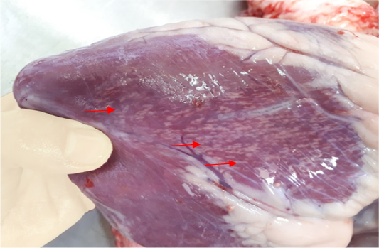
Gross image of calf’s heart shows pathognomic gross lesions termed as “tiger-heart” (arrows).

Microscopic image of calf’s’ heart (Tigroid heart appearance) from calf that died from an acute form of FMD, showing myocarditis represented by focal coagulative necrosis of muscle fibers (arrow), with interstitial infiltration of mononuclear inflammatory cells (arrowhead). (H&E, 100x.).
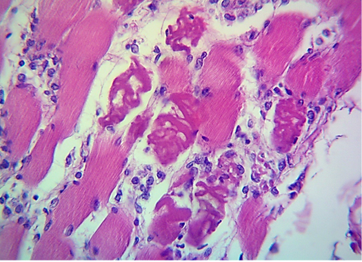
Microscopic image of calf’s’ heart (Tigroid heart appearance) from calf that died from an acute form of FMD, showing focal coagulative necrosis of muscle fibers (arrow) with interstitial infiltration of lymphocytes, plasma cells and histocytes (arrowhead). (H&E, 600x.).
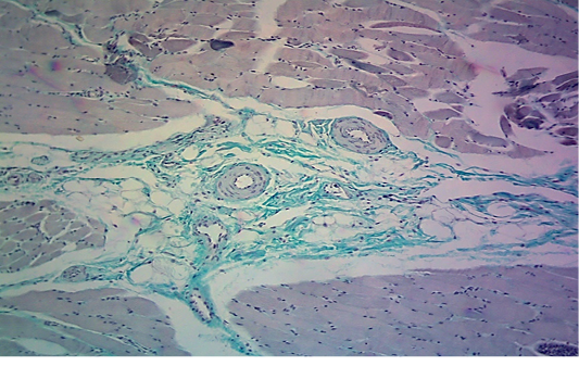
Microscopic image of calf’s heart (“tiger-heart”) from a calf that died from an acute form of FMD, showing interstitial deposition of collagen fibers in green color, with infiltration of mononuclear inflammatory cells in perivascular position (arrow) between collagen fibers. (Masson trichrome stain, 200x.).
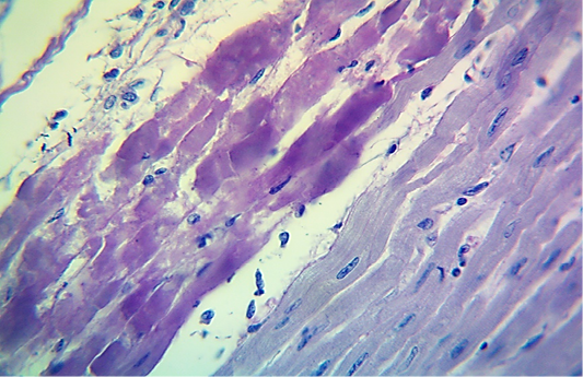
Microscopic image of calf’s heart (“tiger-heart”) from a calf that died from an acute form of FMD, showing severely affected muscle fibers with coagulative necrosis that stained magenta in color (arrow), with infiltration of lymphocytes (arrowhead). (PAS stain, 600x).


