Advances in Animal and Veterinary Sciences
Research Article

Normal Radiographic, Computed Tomography and Ultrasonographic Anatomy of the Carpal Region in Baladi Goats (Capra Hircus)
Attia Ahmed Moselhy1, Mustafa Abd El Raouf2*, Ahmed Mohamed Abdelaal3
1Department of Anatomy and Embryology, Faculty of Veterinary Medicine, Zagazig University, Zagazig - 44519, Egypt; 2Department of Surgery, Anesthesiology and Radiology, Faculty of Veterinary Medicine, Zagazig University, Zagazig, 44519, Egypt; 3Department of Animal Medicine, Faculty of Veterinary Medicine, Zagazig University, Zagazig, 44519, Egypt.
Abstract | Normal diagnostic anatomy of the carpal region of most farm animals was studied, but that of caprine has not been studied in details. Therefore the aim of the present study was to investigate the normal radiography, computed tomography (CT) and ultrasonography of the goat carpal region compared to the dissecting anatomy. Two thoracic limbs were used for carpal bones preparation and examination. In addition, four fresh thoracic limbs separated from the shoulder joint were used for normal anatomical study and latex injection of the joint sacs after CT scanning. The anatomical study was compared with the normal radiography, CT and ultrasonography of the carpal region of five clinically selected healthy goats. Radiographic and CT images revealed normal bone anatomy of the carpal joint, while ultrasonographic imaging revealed ability to visualize tendinous structures of the regional musculature such as those of extensor and flexor muscles as well as joint capsule and collateral ligaments. Therefore, results of the present study were indicated good correlations of radiography, CT and ultrasonography of the goat carpal region with the normal anatomical dissection and provided suitable data for caprine practioners and surgeons as well as educational purposes.
Keywords | Carpal Region, Goat, Radiography, Computed Tomography, Ultrasonography
Received | March 26, 2019; Accepted | May 14, 2019; Published | June 30, 2019
*Correspondence | Mustafa Abd El Raouf, Department of Surgery, Anesthesiology and Radiology, Faculty of Veterinary Medicine, Zagazig University, Zagazig, 44519, Egypt; Email: dr_mustafa13@yahoo.com
Citation | Moselhy AA, Abd El Raouf M, Abdelaal AM (2019). Normal radiographic, computed tomography and ultrasonographic anatomy of the carpal region in baladi goats (capra hircus). Adv. Anim. Vet. Sci. 7(8): 686-693.
DOI | http://dx.doi.org/10.17582/journal.aavs/2019/7.8.686.693
ISSN (Online) | 2307-8316; ISSN (Print) | 2309-3331
Copyright © 2019 Moselhy et al. This is an open access article distributed under the Creative Commons Attribution License, which permits unrestricted use, distribution, and reproduction in any medium, provided the original work is properly cited.
INTRODUCTION
Carpal region is a compound part of the thoracic limb with various structures incorporated carpal joint, ligaments, tendons and muscles (Getty, 1975; Dyce et al., 2009). Carpal joint may be subjected to many pathological changes related to the joint structures, ligaments, tendons of the carpal region (Magnusson and Ekman, 2001; Malone et al., 2003; Vaughan et al., 2014; Jorgensen et al., 2015; Shields et al., 2015).
Radiography and CT is of value in diagnosing musculoskeletal affections and gives accurate morphology of the bones abnormalities (Vallance et al., 2012). On the other hand, it doesnot give accurate diagnosis of the associated soft tissues that are involved (Kofler, 2000). Computed Tomography (CT) is helpful tool for imaging complicated anatomical structures due to its ability to obtain transverse images and manipulate image contrast and latitude (Shojaei et al., 2008).
Ultrasonography is a reliable, rapid, safe, useful, practical diagnostic tool for imaging and evaluating soft tissues and musculoskeletal system and providing valuable information regarding different disorders (Sideri and Tsioli, 2017). Normal ultrasonographic anatomy of the carpal joint of equine (Genovese et al., 1986; Tnibar et al., 1993; Whitecomb, 2014; El-Bably and Abdelgalil, 2018), dogs (Kramer et al., 1997), cattle (Kofler et al., 2014; Al-akraa et al., 2015), camel (Kassab, 2008; Soroori et al., 2011) and sheep (Macrae and Scott, 1999; Sideri and Tsioli, 2017) were studied in details. However, for goats, only ultrasonography of the caprine arthritis which caused by encephalitis virus was described (Moura et al., 2016).
To the author knowledge, very limited studies described normal anatomy of musculoskeletal system in caprine species. Based on current search, there ispoucity of work describing the normal diagnostic anatomy of Baladi goats (Capra Hircus) in Egypt using different diagnostic techniques including radiography, CT and ultrasonography. Therefore, the aim of the current study was conducted to investigate the normal radiography, CT and ultrasonography of the goat carpal region compared to the dissecting anatomy which may help in the diagnosis of different disorders of the carpal region.
MATERIALS AND METHODS
Ethical Approval
All procedures of the current study were approved by and in accordance with the rules of animal use and care ethical committee of Faculty of Veterinary Medicine, Zagazig University, Egypt.
Bone Preparations
Two thoracic limbs were obtained from Zagazig abattoir, then after, the bones of carpal region were prepared by boiling method (Siddiqui et al., 2008). Before boiling, the bones were wrapped separately with a net to prevent the loss of small bones. Prior to boiling, the skin, fascia and muscles were removed from the bones, after that the bones were boiled continuously for three hours. Subsequently, the bones were washed with bleaching solution to remove the unpleasant smell and dried with sunlight for two days.
Computed Tomography And Anatomical Studies
Four fresh thoracic limbs were separated from the shoulder joint and used for normal CT scanning then for anatomical study. The limbs were scanned using CT machine (PHILIPS MX16 EVO CT Scanner, India) with exposure factors 140kv and 256MA. Axial, sagittal and coronal sections were obtained and images were processed using built-in CT software to obtain three dimensional (3D) images. One specimen was used for cross sectional anatomy at three levels (antebrachial, middle carpal and proximal extremity of metacarpus). Findings of the normal anatomical study were compared with the corresponding radiographic, CT and ultrasonographic studies.
Concerning the joint capsule examination, the main compartments of the carpal joint (antebrachiocarpal, middle carpal and carpometacarpal) were injected with gum milk (Latex 60%) colored red with carmine and blue with ultramarine. Latex injection technique was applied on two fresh thoracic limbs. The injected limbs were fixed in a mixture of 10% formalin, 3% glycerine, and 1% thymol. The intrarticular injection was performed at the dorsal aspect and the needle was inserted at the midway between extensor carpi radialis tendon (ECRT) and common digital extensor tendon (CDET). The obtained results were photographed using a Sony digital camera, Dsc W810 20.1 MP.
Radiographic And Ultrasonographic Studies
The radiographic and ultrasonographic studies were carried out on five clinically normal Baladi goats admitted to the clinic of Department of Surgery, Anesthesiology and Radiology – Faculty of Veterinary Medicine – Zagazig University with different ages (1.5-2.5 years) and body weight (35-45 Kg). Dorsopalmar and lateromedial radiographs were performed using X-ray machine (Pox-300 BT, Toshiba, RotanodeTM, Japan) with 45 KV and 6.3 MAs exposure factors. Before ultrasonographic examination, the carpal region from middle forearm region till middle metacarpal region was prepared by hair clipping and shaving for both right and left thoracic limbs. After acoustic coupling gel application, ultrasonographic scanning was performed using a B-mode real-time ultrasound machine (SonoScape A5V, China) connected with 9 MHz linear transducer in a systematic manner of dorsal, medial, palmar and lateral aspects of the carpal joint at longitudinal and transverse scans (El-Bably and Abdelgalil, 2018).
RESULTS
The normal anatomical (Figure 1 & 2), radiographic (Figure 3) and CT (Figure 4) skeleton of the carpal joint was constructed by the distal extremities of radius and ulna, proximal row of carpal bones (radial, intermediate, ulnar, accessory), distal row of carpal bones (fourth and the fused second with third carpal bone) and the proximal extremity of the fused third and fourth metacarpus (large metacarpal bone). The carpal joint is the articulation between antebrachial, carpal and metacarpal bones. It is a compound joint formed from antebrachiocarpal proximally, middle carpal, intercarpal at the middle and carpometacarpal joint distally (Figure 5). Laterally, muscles which associated with the carpal region were common digital extensor muscle, lateral digital extensor muscle, extensor carpi obliques and ulnaris laterals. Whereas, medially the carpal region was associated with deep digital flexor muscle, superficial digital flexor muscle, flexor carpi radialis and flexor carpi ulnaris muscles.
Dorsal Carpal Region
The identified structures on the dorsal aspect of the carpal region were extensor retinaculum, extensor carpi obliqus tendon (ECOT), ECRT and CDET dorsolaterally. Extensor retinaculum was located superficially and covered
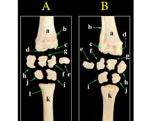
Figure 1: A photograph of bones of the carpal region. (A) Dorsal view, (B) palmar view. a- Radius; b- Ulna; c- Styloid process of ulna; d- Radial articular facets; e- Accessory carpal bone; f- Ulnar carpal bone; g- Intermediate carpal bone; h- Radial carpal bone; i- Fourth carpal bone; j- Fused second and third carpal bone; k- Metacarpal bones; l- Metacarpal tuberosity.
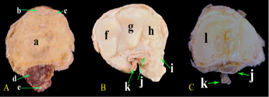
Figure 2: Photomacrograph of the cross sections at different levels of the carpal region. (A) At the distal extremity of radius, (B) At the middle carpal bones, (C) At the metacarpus. a-Radius; b- Extensor carpi radialis M.; c- Common digital extensor M.; d- Deep digital flexor M.; e- Superficial digital flexor M.; f- Radial carpal bone; g- Intermediate carpal bone; h- Ulnar carpal bone; i- Accessory carpal bone; j- Deep digital flexor tendon; k- Superficial digital flexor tendon; l- Metacarpal bones.
by fibrous layer and supporting the joint capsule. It covered and blended with extensor and two collateral ligaments (Figure 6A). It reinforced the extensor aspect of the carpus and fused with the outer fibrous layer of the extensor tendons’ sheath. Ultrasonographically, it appeared as hypoechogenic structure under the skin and above the ECRT (Figure 6C).
The ECRT was existed at the middle dorsal aspect of the carpal region, it was thick, wide, large ribbon-like tendon and it ended at the metacarpal tuberosity (Figure 6B). It was the largest tendon in the carpal region. Ultrasonographically, the ECRT appeared as homogenous echogenic
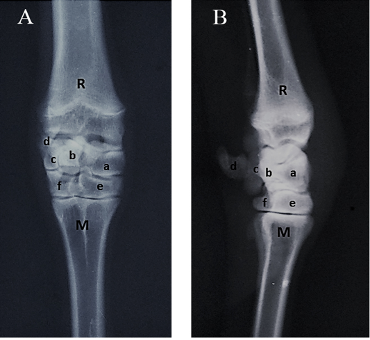
Figure 3: Radiographs of the carpal joint. (A) Dorsopalmar view, (B) Lateromedial view. R- Radius; M- Metacarpus; a- Radial carpal bone; b- Intermediate carpal bone; c- Ulnar carpal bone; d- Accessory carpal bone; e- Fused second, third carpal bone; f- Fourth carpal bone
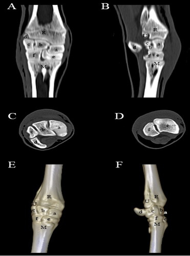
Figure 4: Computed tomography of the carpal joint. (A) Coronal view, (B) Sagittal scanning, (C) Axial scanning of the first raw of the carpal bones, (D) Axial scanning of the second raw of the carpal bones, (E) 3D image of the dorsopalmar aspect of the carpal joint, (F) 3D image of the lateromedial aspect of the carpal joint. R- Radius; U- Ulna; M- Metacarpus; a- Radial carpal bone; b- Intermediate carpal bone; c- Ulnar carpal bone; d- Accessory carpal bone; e- Fused second, third carpal bone; f- Fourth carpal bone.
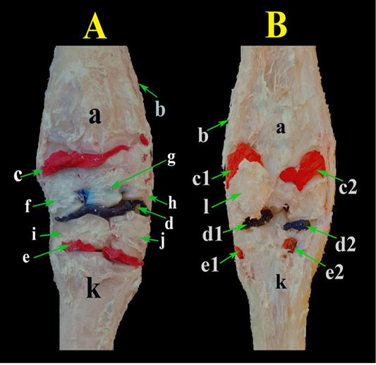
Figure 5: A photograph showing the carpal joint capsule injected with a colored latex. (A) dorsal view and (B) palmar view. a- Radius; b- Ulna; c- Antebrachial-carpal sac; c1- Lateral extension of antebrachial-carpal sac; c2- Medial extension of antebrachial-carpal sac; d- Middle carpal sac; d1- Lateral extention of of middle carpal sac; d2- Medial extension of of middle carpal sac; e- Carpometacarpal sac; e1- Lateral extension of of carpo-metacarpal sac; e2- Medial extension of of carpo-metacarpal sac; f- Radial carpal bone; g- Intermediate carpal bone; h- Ulnar carpal bone; i- Fused second, third carpal bone; j- Fourth carpal bone; k- Metacarpal bones; l- Accessory carpal bone.
structure with parallel linear fibers extends over the carpal joint when scanned longitudinally (Figure 6C). By transverse scanning, it appeared as an oval echogenic structure with anechoic fluid within tendon sheath when scanned at the level of distal extremity of the radius and antebrachiocarpal joint (Figure 6D).
The joint capsule (JC) of carpal joint found as a fibrous sheet covered externally all structures of the joint, it lodged the distal end of antebrachial bones proximally and the proximal end of the metacarpus distally. This capsule was closely adherent to the middle carpal ligament and the carpal bones (Figure 6B). By ultrasonographic examination, the JC appeared as hypoechoic structure beneath ECR tendon with anechoic synovial fluid. The antebrachiocarpal joint sac was identified clearly in flexed position but the middle carpal and carpometacarpal joint sacs were hardly identified neither in extended nor flexed positions (Figure 6C).
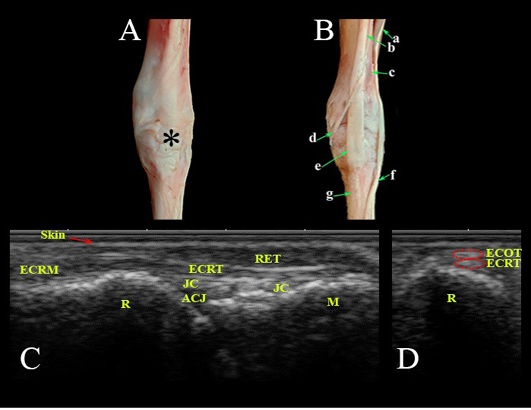
Figure 6: Dorsal aspect of the carpal region. Photomacrographs: (A) superficial layer and (B) deep layer. Ultrasonographic imaging: (C) Longitudianl scan (LS) and (D) Transverse scan (TS) at the level of the radius. *- Extensor retinaculum; a- Common digital extensor M.; b- Extensor carpi radialis M.; c- Extensor carpi obliqus M.; d- Tendon of extensor carpi obliqus M.; e- Tendon of extensor carpi radialis; f- Tendon of common digital extensor M.; g- Metacarpal bones; Ret- Retinaculum; ECRT- Extensor carpi radialis tendon; ECRM- Extensor carpi radialis M.; ECOT- Extensor carpi obliqus tendon; JC- Joint capsule; ACJ- Antebrachiocarpal joint; R- Radius; M- Metacarpus.
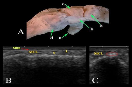
Figure 7: Medial collateral ligament of the carpal joint. (A) Photomacrograph. Ultrasonographic imaging: (B) LS, (C) TS. a- Radius; b- Medial collateral ligament; c- Accessory carpal bone; d- Metacarpal bones; (e) Radial carpal bone; MCL- Medial collateral ligament; L- Long fibers; S- Small fibers.
Medial Carpal Region
This region contained, the ECOT, which passed transversely from the distal part of the radius laterally to be inserted at the mediopalmar aspect of the proximal extremity of the base of the large metacarpal bone. This tendon was long, slender and crossed obliquely above the ECRT (Figure 6B). Ultrasonographically, ECOT appeared as an oval echogenic structure above ECRT when examined longitudinally and transversally at the level of distal radius (Figure 6D).
The medial collateral ligament (MCL) was long continuous structure, extended from the distal extremity of the radius and inserted at the medial aspect of the proximal extremity of the large metacarpal bone. Its insertion point had two branches, long and short fibers. The long fibers attached to the large metacarpal bone, while the short fibers attached to the radial carpal and second carpal bones (Figure 7A). It appeared ultrasonographically as echogenic band with parallel pattern in longitudinal scan at the distal radius and as irregular fibers at the distal half with easily identification of short and long fibers (Figure 7B). While in transverse scan, it appeared as flattened oval echogenic structure (Figure 7C).
Palmar Carpal Region
There were several structures present in this area including the deep digital flexor (DDF) muscle (humeral, radial, ulnar heads), and its tendon and superficial digital flexor (SDF) muscle (superficial, deep bellies) with its tendon (Figure 8A).
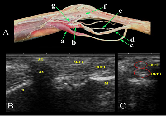
Figure 8: Palmar aspect of the carpal region. (A) Photomacrograph of the deep dissection of the forearm region and carpal canal. Ultrasonographic imaging: (B) LS and (C) TS. a- Superficial digital flexor M.; b- Deep digital flexor M.; c- Superficial digital flexor tendon; d- Deep digital flexor tendon; e- Median Nerve.; f- Carpal canal opened; g- Median Artery; AC- accessory carpal bone; AS- Acoustic shadow; SDFT- Superficial digital flexor tendon; DDFT- Deep digital flexor tendon; R- Radius; M- Metacarpus.
In this region, the carpal canal was found. It was bounded laterally by accessory carpal bone, dorsally by palmar carpal ligament and mediopalmarly by medial collateral ligament and flexor retinaculum (transverse palmar carpal ligament) which completely closed the carpal canal. The canal was filled by SDFT and DDFT, as well as median artery and nerve (Figure 8A). Ultrasonographic examination of SDF muscle revealed a heterogenous echogenic structure with intermeshed anechoic and echogenic bands. The DDF muscle appeared as a heterogenous echogenic structure with its four heads. The SDF and DDF tendons appeared as echogenic structures with homogenous parallel tendon fibers and DDFT was more echogenic than SDFT (Figure 8B). The median vein and artery appeared as an oval anechoic structure, while the median nerve appeared as a round hypoechogenic structure.
Lateral Carpal Region
The most prominent structure in this area was the lateral collateral ligament (LCL). It started from the distal extremity (styloid process) of the ulna and extended toward the accessory and ulnar carpal bones and to the lateral part of the proximal extremity of the metacarpal bone (Figure 9A). It appeared ultrasonographically as a thin echogenic band in longitudinal scan and as a small flattened structure in transverse scan (Figure 9B).
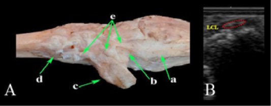
Figure 9: Lateral collateral ligament of the carpal joint. (A) Photomacrograph and (B) TS Ultrasonographic imaging. a- Ulna; b- Ulnar carpal bone; c- Accessory carpal bone; d- Metacarpal bones; e- Lateral collateral ligament; LCL- Lateral collateral ligament.
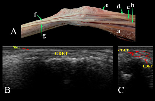
Figure 10: Lateral aspect of the carpal region. (A) Photomacrograph. Ultrasonographic imaging of the dorsolateral aspect of the carpal region: (B) LS and (C) TS. a- Ulnaris lateralis; b- Lateral digital extensor M.; c- Common digital extensor M. lateral belly; d- Common digital extensor M., medial belly; e- Extensor carpi obliqus M.; f- Tendon of common digital extensor; g- Tendon of lateral digital extensor M; LDET- Lateral digital extensor tendon; CDET- Common digital extensor tendon.
The CDE and lateral digital extensor (LDE) tendons were situated at the dorsolateral aspect of the carpal region. The CDET was divided into two parts, medial and lateral, The medial one was larger (Figure 10A). They appeared ultrasonographically as echogenic bands in longitudinal scan (Figure 10B) and as oval structures in transverse scan (Figure 10C).
Special Ligaments of the Carpal Joint
The dorsal radiocarpal ligament extended from the distal extremity of the radius to intermediate and ulnar carpal bone. The dorsal intercarpal ligament passed from radial carpal bone to fourth carpal bone. The dorsal carpometacarpal ligament passed from fourth carpal bone to the proximal extremity of the metacarpal (Figure 11A). The palmar radiocarpal ligament was short ligament passed from the distal extremity of the radius at the palmar aspect of the ulnar carpal bone. The palmar ulnocarpal ligament was long ligament connected the distal extremity of the ulna to the radial carpal bone. The palmar carpometacarpal ligament passed from the radial carpal bone to the proximal extremity of the metacarpal (Figure 11B & C).
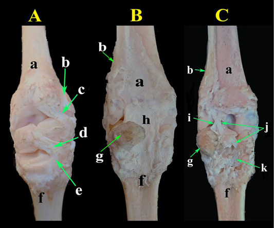
Figure 11: Photomacrographs of the special ligaments of the carpal joint. (A) Dorsal view, (B) Palmar view and (C) Palmar view after removal of the common palmar carpal ligament. a- Radius; b- Ulna; c- Dorsal radiocarpal ligament; d- Dorsal intercarpal ligament; e- Dorsal carpometacarpal ligament; f- Metacarpal bones; g- Accessory carpal bone; h- Common palmar carpal ligament; i- Palmar radiocarpal ligament; j- Palmar ulnocarpal ligament; k- Palmar carpometacarpal ligament.
DISCUSSION
Normal anatomy of the carpal region and its related soft tissue structures were described in previous studies for horses (Tnibar et al., 1993; Whitcomb, 2008, 2014; El-Bably and Abdelgalil, 2018), cattle (Kofler, 2000; Al-akraa et al., 2015), camels (Kassab, 2008; Soroori et al., 2011), dogs (Kramer et al., 1997), and sheep (Macrae and Scott, 1999; Sideri and Tsioli, 2017), while the present study gives a complete description of the bones, tendons, ligaments, and other soft tissue structures of the carpal region of goats during their course over the carpus.
The number and arrangement of bones of the goat carpal joint were similar in dissecting, radiographic and CT investigations. Only the large metacarpal bone was present, which formed from the fusion of third and fourth metacarpal bones and other metacarpal bones were absent. In addition to extensor and flexor muscles, tendons, ligaments, and carpal joint sacs and capsule. These findings were in agreement with those reported in textbooks (Getty, 1975; Nickel et al., 1986; Siddiqui et al., 2008; Hussain, 2010).
Ultrasonography is a non-invasive, economic, non-irritant, rapid and easily applied diagnostic technique and used currently for diagnosis and evaluation of musculoskeletal disorders of animals (Soroori et al., 2011).
In the current work, a 9 MHz linear transducer provided better resolution and imaging for all structures of the carpal region that nearly used for ultrasonography of the carpal joint in other different animals including horses (Tnibar et al., 1993; El-Bably and Abdelgalil, 2018), cattle (Al-akraa et al., 2015), camel (Kassab, 2008; Soroori et al., 2011), dogs (Kramer et al., 1997) and sheep (Macrae and Scott, 1999; Sideri and Tsioli, 2017). The transverse scanning of the extensor tendons has better advantage than longitudinal scanning as they were side by side at their course and could not be distinguished longitudinally. Ultrasonography of the tendons and ligaments of the carpal region was performed separately either longitudinally or transvesly. These findings were similar to those of horses (Tnibar et al., 1993; El-Bably and Abdelgalil, 2018) and cattle (Kofler, 2000; Al-akraa et al., 2015) and in contrast to lower limb ultrasonography (metcarpal, metatarsal and pastern regions) examination where the tendons could be scanned at a single acoustic window (Genovese et al., 1986; Kofler, 1995).
Ultrasonographic appearance of the longitudinal and transverse scans of the tendons had an even echogenic structure, while at the muscle-tendon transition it varied from hypoechogenic to echogenic structure. These results were in agreement with ultrasonographic appearance of tendons in horse (Genovese et al., 1986; Tnibar et al., 1993; Whitecomb, 2014; El-Bably and Abdelgalil, 2018).
The ultrasonographic images of the extensor and flexor tendons were clear when they examined over the radius, the carpus and at the level of proximal metcarpus. The dissecting anatomy of the carpal structures was well correlated to the ultrasonographic images. The ultrasonography of ECR, CDE, SDF and DDF tendons was clear and easily identified from the neighbouring structures.
The tendon of ECO muscle was visualized ultrasonographically over the radius while it crossing the tendon of ECR muscle. The JC can be imaged as hypoechoic structure that might be attributed to the fat cushion present under the ECRT. The synovial fluid in the antebrachiocarpal joint sac could be visualized as anechoic fluid especialy in semiflexed position of the limb, while the middle carpal and carpometacarpal joint sacs could not be identified ultrasonographically. Similar observations were reported in horses (Whitcomb, 2008; Whitecomb, 2014; El-Bably and Abdelgalil, 2018). These structures were more easily identified in diseased cases (Penninck et al., 1990; Reed et al., 1995; Kofler, 2000; Moura et al., 2016).
The MCL was larger than the LCL and irregular fiber patterns were detected at its distal half toward the metacarpal insertion that might be attributed to the different fiber orientation at their insertion. Similar findings were reported in horses (Whitcomb, 2008; El-Bably and Abdelgalil, 2018).
The fleshy part of SDF and DDF tendons proximal to the accessory carpal bone that appeared as echogenic line with distal acoustic shadowing appeared as a heterogenous echogenic with anechoic areas. These findings were in line with (Whitecomb, 2014; El-Bably and Abdelgalil, 2018) in horses. The SDF and DDF tendons distal to the accessory carpal bone had similar echogenicity and the DDF tendon was more echogenic sometimes. These findings were in agreement with those observations in horses (Cuesta et al., 1995), cattle (Kofler, 2000; Al-akraa et al., 2015) and camel (Kassab, 2008).
CONCLUSIONS
Radiographic, CT and ultrasonographic anatomy of the goat carpal region were in correspondence with dissecting anatomy that generate a valuable information essential for clinical and surgical practices.
ACKNOWLEDGEMENTS
The authors would like to thank Department of Surgery, Anatomy and Animal Medicine for their support and providing all facilities for this work. The authors did not get any fund for this work.
CONFLICT OF INTEREST
The authors declare that they have not conflict of interest.
AUTHORS’ CONTRIBUTIONS
All auhtors contribute to this research equally. They made available relevant literatures and conducted examinations as well. All authors participated in draft and revision of the manuscript. All authors read and approved the final manuscript.
REFERENCES


