Advances in Animal and Veterinary Sciences
Research Article

Clinical and Ultrasonographic Observations of Omental Affections as Uncommon Causes of Mechanical Ileus in Cattle
Ahmed M. Abdelaal1*, Ahmed Abdelbaset-Ismail2*, Shaimaa M. Gouda1, Mohamed Gomaa2, Mohamed Aref3, Hassan Emam3, Kuldeep Dhama4, Ahmed Ezzat Ahmed5,6
1Department of Animal Medicine, Faculty of Veterinary Medicine, Zagazig University, 44519 Zagazig, El-Sharkia, Egypt; 2Department of Surgery, Anesthesiology and Radiology, Faculty of Veterinary Medicine, Zagazig University, 44519, Zagazig, El-Sharkia, Egypt; 3Department of Anatomy and Embryology, Faculty of Veterinary Medicine, Zagazig University, 44519 Zagazig, El-Sharkia, Egypt; 4Division of Pathology, ICAR-Indian Veterinary Research Institute, Izatnagar, India; 5Biology Department, College of Science, King Khalid University, 61413 Abba, Kingdom of Saudi Arabia; 6Theriogenology Department, Faculty of Veterinary Medicine, South Valley University, 83523 Qena, Egypt.
Abstract | Mechanical ileus as the major cause of intestinal obstruction in cattle can arise from usual cases including complicated hernia, volvulus and intussusception. Our previous study reported some unusual cases causing ileus such as diaphragmatic hernia and internal localized abscessation in bovines. The purpose of this study was to define the diagnostic features and to show the representative images of other uncommon cases of mechanical ileus caused by omentum disorders in cattle. One-hundred-sixty-five cattle were referred to Veterinary Clinic of Zagazig University with abdominal distension and signs of ileus for clinical and abdominal ultrasound examinations. The anatomical and necropsy examinations were additionally involved in this study. Herein, we present uncommon cases of omentum- derived mechanical ileus in cattle. Among 165 total cases, ten cases (~6.1%) were identified as omentum disorders. More interestingly, as shown by the animal records, these ten animals underwent right omentopexy operation for correction of left displaced abomasum (LDA), 25±5 days pre- admission to the clinic. The following cases were reported: omental hernia (OH; 3 out of 10), omental fat necrosis (OFN; 2 out of 10) and omental bursitis (OB; 5 out 10). The observed necropsy findings confirmed the tentative diagnosis that was reached upon the ultrasonographic data. Based on the data presented here, omental affections should be involved during examination of cattle with mechanical ileus secondary to LDA surgical corrections.
Keywords | Abdominal distension, Cattle, Omentum, Ileus, Ultrasound
Received | March 04, 2018; Accepted | April 11, 2019; Published | May 26, 2019
*Correspondence | Ahmed M Abdelaal, Department of Animal Medicine, Faculty of Veterinary Medicine, Zagazig University, 44519, Egypt; Ahmed Abdelbaset- Ismail. Department of Surgery, Anesthesiology and Radiology, Faculty of Veterinary Medicine, Zagazig University, 44519, Egypt; Email: ahmed.abdelaal@zu.edu.eg; a4azzazy@yahoo.com
Citation | Abdelaal AM, Abdelbaset-Ismail A, Gouda SM, Gomaa M, Aref M, Emam H, Dhama K, Ahmed AE (2019). Clinical and ultrasonographic observations of omental affections as uncommon causes of mechanical ileus in cattle. Adv. Anim. Vet. Sci. 7(7): 543-549.
DOI | http://dx.doi.org/10.17582/journal.aavs/2019/7.7.543.549
ISSN (Online) | 2307-8316; ISSN (Print) | 2309-3331
Copyright © 2019 Abdelaal et al. This is an open access article distributed under the Creative Commons Attribution License, which permits unrestricted use, distribution, and reproduction in any medium, provided the original work is properly cited.
INTRODUCTION
Ileus that is not cured by classical medical treatment is a great irritant to both clinicians and animals’ owners due to its confusing symptoms and substantial economic burden, respectively (Braun et al., 1993). The most important type of ileus that always leads to intestinal obstruction is mechanical-paralytic ileus (Pardon et al., 2009; Ruf-Ritz et al., 2013). In spite of full clinical examination as well as biochemical and hematological analysis of blood and peritoneum fluid are routinely performed in such cases, however, their proper diagnosis and prognosis are still being equivocal (Pardon et al., 2009).
Omentum as a valuable component of abdomen could be affected and in turn shares in progress of ileus in cattle. Mounting evidences have indicated that the omentum occupies a central position in the peritoneal defense system (Platell et al., 2000). It fulfills this function through immune aggregates located in its perivascular areas “milky spots”, absorptive capabilities of germs and other contaminants, providing a layer of fibrin that consequently form collagen wall bounding and adhering the site of inflammation (Shimotsuma et al., 1993). Another well-known function of omentum is to maintain the intestine warm (Hall et al., 1998; Platell et al., 2000).
Despite these various advantages, the omentum may act as a home of some rare pathological changes (Schaudien et al., 2007). There are so far scarce case reports that have reported cases of omental disorders in cattle (Hekmati and Zakarian 1971; van Beukelen et al., 1979; Grymer et al., 1982; Pardon et al., 2009; Tharwat and Buczinski 2012). These reports, however, depend mainly on necropsy to reach diagnosis of such kind of cases. The experiencing clinical findings of the omentum–derived ileus are not actually specific. Hence, as in the previous, the exploratory laparotomy is only way for reaching the ultimate diagnosis of these uncommon disorders and consequently having a proper judgment. Additionally, most of such cases are ultimately referred to the hospital for addressing the problem by an expert sonographer. Thus, as previously reported, ultrasonographic examination of the abdomen plays a central role in diagnosis of internal abdominal abscesses (Abdelaal et al., 2017), diaphragmatic hernia (Abdelaal et al., 2014) and omental hernia (Pardon et al., 2009) in bovines.
In order to expand our knowledge at this important area, this study therefore presents some rare cases of mechanical-paralytic ileus that primarily due to omental disorders in cattle according to the confirmed data obtained by ultrasonography and postmortem examination.
MATERIALS AND METHODS
All institutional and national guidelines for the care and use of animals were followed according to the Egyptian Medical Research Ethics Committee.
Animals
Ten cases were selected from 165 cattle suffering from abdominal distension and general signs of ileus that were refereed to Veterinary Clinic of Zagazig University between December 2016 and October 2018. All animals were female non-pregnant with age range 3 to 7 years and weighed 450- 650 kg. Based on ultrasonography and necropsy findings, these cases were identified as omental hernia OH (n=3),omental fat necrosis OFN (n=2) and Omental bursitis OB (n=5). Additionally, 5 apparently healthy non-pregnant cattle of the same age and weight were also included as a control group.
History and Clinical Examinations
Three days before admission, these animals were received a symptomatic treatment by field veterinarians. Access to the records of these cattle revealed that they underwent right omentopexy operation for correction of LDA 25±5 days pre-admission to the clinic. Owing to the unresponsive treatment, these animals were referred to our clinic. On presentation, rectal temperature, heart rate, respiratory rate were recorded. Further, ruminal contractions, abdominal auscultation and ballottement, abdominal circumferences and rectal palpation were performed and their data were recorded. All examinations were performed by the methods described previously (Rosenberger 1990).
Ultrasonographic Examination and Anatomical Schematic Representation
Abdominal ultrasonographic scanning was performed per each animals (control and diseased groups). Six topographic areas were selected to assess the different abdominal organs. The first was at ventral abdominal wall between xiphoid and umbilicus to assess reticulum in relation to rumen and abomasum, 2nd was at right ventral abdomen at the level of elbow to assess reticulum in relation to the liver, 3rd at right flank to asses small and large intestine, 4th at right last 3 intercostal spaces to assess liver and gallbladder, the 5th was at left flank to assess rumen and check any abnormalities and 6th was at left ventral abdomen at the level of elbow to assess the reticulum in relation to the spleen. All selected areas were shaved and coupling gel was applied before examination. A 3.5- 5 MHz convex probe ultrasound was used (Sono Scape A5V ultrasound machine, China). This examination was done as previously outlined (Braun 2003).
For better understanding, the anatomical schematic drawing of omentum in control group at the level of 4th lumbar and 13th thoracic vertebrae, in addition to the illustrations of different sites where lesions were observed in diseased cattle were accomplished using Adobe Photoshop CC program.
Necropsy Examination
Out of examined cases, 6 cases (2 for each) were followed after leaving the clinic in order to properly confirm the tentative ultrasonographic diagnosis by postmortem investigation.
RESULTS
Clinical Findings of Cattle with Omental Affections
As shown in Table-1, Anorexia, abdominal distension, sh-
Table 1: Frequency distribution (n) and percentage (%) of clinical findings in 10 diseased cattle in comparison to 5 controlnormal cattle.
|
Clinical findings |
Control (n=5)
|
Omental affections (n=10) | |||
| OH (n=3) | OFN (n=2) | OB (n=5) | Total N (%) | ||
| Anorexia | 0 | 3 | 2 | 5 | 10 (100) |
| Abdominal distension (right ventral) | 0 | 3 | 2 | 3 | 8 (80) |
| Abdominal distension (papple abdomen) | 0 | 0 | 0 | 2 | 2 (20) |
| Scanty feces | 0 | 3 | 2 | 5 | 10 (100) |
| Abdominal pain | 0 | 3 | 2 | 2 | 7 (70) |
| Dehydration (mild) | 0 | 3 | 2 | 5 | 10 (100) |
| Fluid splashing (abdominal agitation) | 0 | 3 | 2 | 5 | 10 (100) |
|
Melena |
0 | 0 | 0 | 2 | 2 (20) |
| Systemic reactions* | 0 | 1 | 0 | 5 | 6 (60) |
| Congested mucous membranes | 0 | 1 | 1 | 2 | 4 (40) |
| Decrease in milk production | 0 | 3 | 2 | 5 | 10 (100) |
|
Ruminal movements/ 2 minutes 2-4 0-2 |
5 0 |
0 3 |
0 2 |
0 5 |
0 (0) 10 (100) |
| Empty rectum by rectal palpation | 0 | 3 | 2 | 5 |
10 (100) |
*Systemic reactions include rising of body temperature above 38.8±0.2oC, respiratory rate above 25.6±6.2/minute and heart rate above 74±5.8/minute.
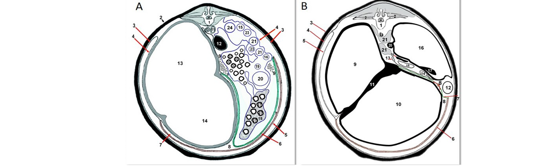
Figure 1: A. Normal schematic diagram of normal omentum and omental bursa of cattle at the level of 4th lumbar vertebra. 1) Fourth lumbar vertebrae; 2) Abdominal wall; 3) Parietal peritoneum; 4) Visceral peritoneum; 5) Superficial layer of greater omentum; 6) Deep layer of greater omentum; 7) Omental bursa; 8) Caudal omental recess; 9) supraomental recesses; 10) Abdominal aorta; 11) Caudal vena cave; 12) Left kidney; 13) Dorsal ruminal sac; 14) Ventral ruminal sac; 15) Ascending duodenum; 16) Descending duodenum; 17) Jejunum; 18)
arp decrease in milk production, hypo-motile rumen, mild degrees of dehydration, and scanty feces either hard or soft were the main signs and appeared in all affected cases (n= 10; 100%). Abdominal circumference revealed distension of right ventral abdomen in eight cases, and papple shape in the remaining two cases of OB. Fluid splashing sound, as predominant recorded sign, was detected by abdominal ballottement of ventral abdomen. Melena or dark tarry
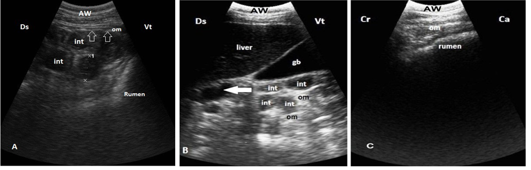
Figure 2: Ultrasonogram shows normal moderate echogenicomentum. A. The intestinal loops (int) are easily visible between rumen and right abdomen from right ventral flank and covered by omentum (arrows). B. Omentum covering the intestinal loops (int) medial to the liver and gallbladder (GB) at 10th ICS, note that duodenum (arrow) not covered by omentum. C.Omentum covers the whole ventral sac of the rumen, imaged from ventral abdomen. Ds: dorsal; Vt: ventral; Cr: cranial; Ca: caudal
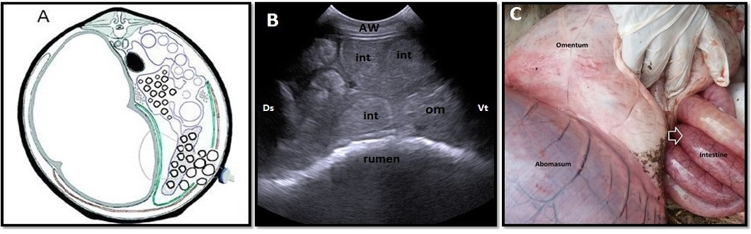
Figure 3: Omental herniation “entrapment” of 3-years-old-cow. A. Cross-section schematic drawing of abdomen at the level of 4th lumbar vertebra. Note intestinal entrapment through torn greater omentum. B. Ultrasonogram of ventral part of right flank region with distended intestinal loops located beneath abdominal wall with ruptured omentum (om), which located medial to intestineC. Lesions observed at necropsy. Note congestion and distension of entrapped intestinal parts through torn omentum (arrow). AW: abdominal wall; Ds: dorsal; Vt: ventral; Cr: cranial; Ca: caudal
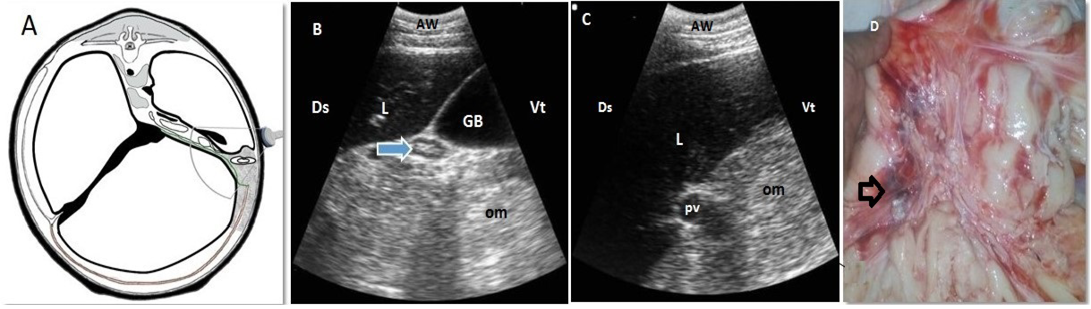
Figure 4: Omental fat necrosis “lipomatosis” of 3-years and 6-months-old-cow. A. Cross-section schematic drawing of abdomen at the level of 13th thoracic vertebra. Note the location of the disease and the transducer. B, C. Ultrasonogram of ventral part of right 10th intercostal space. Note presence of echogenic masses within the omentum (om) with invisible intestinal loops and a compressed duodenum (arrow). D. Lesions presented at necropsy. Note the reddish and blackish discoloured omentum fatty tissues (arrow). AW: abdominal wall; L:liver; GB: Gallbladder; pv: portal vein, Ds: dorsal; Vt: ventral
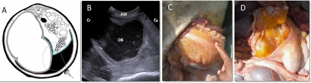
Figure 5: Omental bursitis of 4-years-old-cow. A. Cross-section schematic diagram of abdomen at the level of 4th lumbar vertebra. Note the site of inflamed bursa and the location of transducer. B. Ultrasonogram of the same location showing an extensive collection of anechoic exudate with echogenic debris filling the space between superficial and deep layers of greater omentum. C, D. Lesions noticed at necropsy. Note presence of enlarged sac “omental bursa” containing yellowish, gelatinous exudate. AW: abdominal wall; Cr: cranial; Ca: caudal; OB: omental bursa
feces was observed in two cases of OB. Pain reactions including arched back, abducted elbows and expiratory grunting were exhibited by the most of diseased cases. All cases of OB showed disturbances in the body temperature, heart rate and respiratory rate. Petechial hemorrhage on vulvar and conjunctiva mucous membrane and engorged eye capillaries were other observed signs
Anatomical Schematic Representation of Normal Omentum
For better understanding, the anatomical schematic drawing of normal double layers of greater omentum (superficial and deep layers) covering the ventral ruminal sac and intestinal loops and omental bursa of cattle at the level of 4th lumbar and 13th thoracic vertebrae are depicted in Figure-1 A and B.
Ultrasonographic Findings
In control animals, the omentum appeared as a moderate
echogenic structure covering the intestinal loops (with exception the cranial duodenum in case of right 10th ICS scanning) (Figure 2A and B). Based on scanning site, the omentum was visualized medial to the abdominal wall or to the liver when scanned at right ventral flank (Figure 2A) or at right 10th ICS (Figure 2B), respectively. At both scanning sites, the intestinal loops were easily visualized with diameter of 2.5±1 cm. Other than the reticulum and abomasum, the omentum appeared covering only the whole ventral ruminal sac (Figure. 2C) as indicated by another scanning approach through ventral abdomen.
Omental hernia appeared at right abdominal wall. Distended intestinal loops located just medial to intact abdominal wall and uncovered by omentum. The omentum in these cases appeared as moderate echogenic mass and located medial to the intestine (Figure 3A and B). Omental fat necrosis was detected at right abdominal wall medial to the liver as hyperechoic structures that completely affected the clear visibility of the intestinal loops. Empty duodenum was observed in only one case of OFN (Fig. 4 A,B, and C). Omental bursitis was visualized at ventral abdominal wall as echogenic-circumscribed wall with anechoic content and echogenic debris (Figure 5A and B).
Necropsy Findings
As inspected, the results of postmortem verification were consistent with ultrasound data. In cases of OH, hemorrhagic and distended intestinal loops were evident uncovered with omentum directly under the parietal layer of peritoneum at the area where localized double layers of greater omentum were ruptured (Figure 3C). While in cases of OFN, the omentum appeared hard and thick with various focal hemorrhagic spots (Figure 4D). In case of OB, the inflamed omental bursa was directly found between xiphoid and umbilicus after incising the ventral abdominal wall due to accumulation of exudate between superficial and deep layer of greater omentum inside the bursa at the ventral abdominal wall. After the incision of the exposed omental bursa, yellowish-gelatinous exudate was drained out indicating much fibrin in exudate (Figure 5C and D).
DISCUSSION
It is well known that omentum-derived ileus is rare and always confused with other ileus leading disorders in cattle. This kind of ileus may occur due to OH, OFN (or lipomatosis), or OB (Hekmati and Zakarian 1971; Grymer et al., 1982; Pardon et al., 2009; Tharwat and Buczinski 2012). All these forms are clinically accompanied by abdominal distension, so that, it is necessary to be critically diagnosed and differentiated from other disorders causing abdominal distension (for example: intestinal invagination and twisting as well as abomasal displacement) (Roussel et al., 2000; Lejeune et al., 2015). As well, it is not possible to clinically diagnose such rare omental disorders because of their non-specific experiencing symptoms including anorexia, dehydration, abdominal pain, and systemic disturbances. By this study, the experiencing signs including lack of appetite and demeanor, abdominal pain, elevated respiration and pulse, scanty hard feces were recorded in animals affected with OB (Hekmati and Zakarian 1971; van Beukelen et al., 1979). Other specific sign such as melena and papple abdomen that observed only in 2 cases of OB could indicate that the origin of this disease is perforated abomasal ulcer with secondary vagal indigestion (Constable et al., 2016).
Based on this, grasping the exact diagnosis of these uncommon diseases necessitates employing other diagnostic methods that should be non-invasive, inexpensive, and practically available. Ultrasonographically is the best instance because it helps not only in diagnosis but also in determination of nature and extent of the illness-causing lesions (Braun et al., 1995; Seyrek-Intas et al., 2005). At this regards, our previous reports have evidenced that the ultrasonography is an important tool in diagnosis and characterization of the diaphragmatic hernias (Abdelaal et al., 2014), and various kinds of internal abscessations in cattle and buffaloes (Abdelaal et al., 2017).
In the current study, the fat of the greater omentum in normal animals was not hard, therefore, it appeared with moderate echogenicity and the underlying organs could be visualized. The greater omentum has a high value in protection of entire organs and appeared medial to the right abdominal wall. The intestinal loops were clearly appeared with the diameter of 2.5±1 cm and covered by a moderate echogenic omentum with the exception of duodenum. This result is consistent with the previously reported data (Braun 2003). Moreover, the data shown here also provides an additional support of such unique role of ultrasonography involving potential assistance in diagnosis and characterization of three uncommon forms of omental diseases secondary to right ometopexy of LDA.
Omental hernia where a piece of intestine was found prolapsed and entrapped medial to abdominal wall through a local tear of omentum (Pardon et al., 2009; Ruf-Ritz et al., 2013). This entrapment consequently disturbs the intestinal motility and compresses the intestine leading to distension of affected loops (5±0.5 cm diameter) mostly at right abdominal wall (Pardon et al., 2009) that were also demonstrated herein by ultrasonography and then confirmed by necropsy. Another recorded affection in this study is OFN (or abdominal/omental lipomatosis) was successfully diagnosed. As evident by several reports, omental lipomatosis is a pathological process mainly occurred in adipose tissue of omentum with unknown etiology (Linnenkohl et al., 2013). Also, at the past, this kind of disorders was only discovered at slaughterhouse (Aydın 1995). Whereas, as we show here, the omental lipomatosis was diagnosed in three cases with hyperechoic structures medial to the liver at the right abdomen where the visibility of intestine was completely hindered. Additionally, hard thick lipomatous omentum with various focal hemorrhagic spots was also noticed at necropsy. Additionally, OB was detected upon ultrasonographic examination as large sac at ventral abdominal wall that bounded by echogenic wall of varying thickness and anechoic contentand echogenic debris.Omental bursitis is a local form of peritonitis characterized by collection of inflammatory exudate within the so-called omental bursa (Grymer et al., 1982). These cases were afterwards confirmed by postmortem examination that indicated presence of swollen omental bursa filled with yellowish-gelatinous exudate at the area between xiphoid and navel. This kind of cases could clinically be differentiated from diffused peritonitis in cattle by right-side involvement, and severe loss of demeanor and appetite, and systemic disturbances in case of the latter case (Abdelaal et al., 2009).
Conclusion
Thorough assessment of cattle cases having mechanical ileus is important to fully figure out the exact diagnosis and consequently the prognosis, since there are various abdominal disorders that are uncommonly occurred. Omentum is considered as a home of such uncommon pathological disorders that actively share in progression of mechanical ileus. As extracted from this study, ultrasonography could be potentially used for grasping the exact diagnosis of these disorders in cattle especially for that of surgical history. Taken all, the omentum should be spotted during examination of animals having ileus and abdominal distension after a period of abdominal surgery.
ACKNOWLEDGEMENTS
This work was supported by Faculty of Veterinary Medicine, Zagazig University, Egypt. Special thanks to Vet. Dr./ Mohamed Gharib for his help in this study.
CONFLICTS OF INTEREST
The authors declare that there are no conflicts of interest.
authors contribution
This study was fulfilled by a consistent contribution of the all listed authors.
REFERENCES


