Advances in Animal and Veterinary Sciences
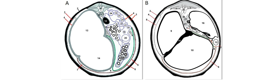
A. Normal schematic diagram of normal omentum and omental bursa of cattle at the level of 4th lumbar vertebra. 1) Fourth lumbar vertebrae; 2) Abdominal wall; 3) Parietal peritoneum; 4) Visceral peritoneum; 5) Superficial layer of greater omentum; 6) Deep layer of greater omentum; 7) Omental bursa; 8) Caudal omental recess; 9) supraomental recesses; 10) Abdominal aorta; 11) Caudal vena cave; 12) Left kidney; 13) Dorsal ruminal sac; 14) Ventral ruminal sac; 15) Ascending duodenum; 16) Descending duodenum; 17) Jejunum; 18) Mesojejunum; 19) Ileum; 20) Caecum; 21) Proximal loops of ascending colon; 22) Spiral loops of ascending colon; 23) Distal loops of ascending colon; 24) Descending colon. B. Normal schematic diagram of normal omentum and omental bursa of cattle at the level of 13th thoracic vertebra. 1) Thirteenth thoracic vertebra; 2) Thirteenth rib; 3) Abdominal wall; 4) Parietal peritoneum; 5) Visceral peritoneum; 6) Superficial layer of greater omentum; 7) Deep layer of greater omentum; 8) Omental bursa; 9) Dorsal ruminal sac; 10) Ventral ruminal sac; 11) Right ruminal pillar; 12) Ascending duodenum; 13) Descending duodenum; 14) Jejunum; 15) Colon; 16) Liver; 17) Gallbladder; 18) Portal vein; 19) Aorta; 20) Caudal vena cava; 21) Crura of diaphragm.
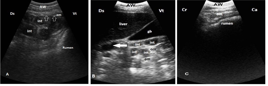
Ultrasonogram shows normal moderate echogenicomentum. A. The intestinal loops (int) are easily visible between rumen and right abdomen from right ventral flank and covered by omentum (arrows). B. Omentum covering the intestinal loops (int) medial to the liver and gallbladder (GB) at 10th ICS, note that duodenum (arrow) not covered by omentum. C.Omentum covers the whole ventral sac of the rumen, imaged from ventral abdomen. Ds: dorsal; Vt: ventral; Cr: cranial; Ca: caudal
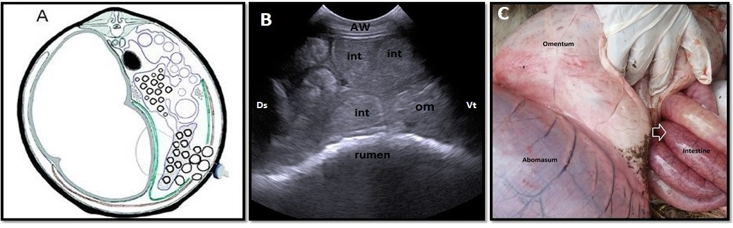
Omental herniation “entrapment” of 3-years-old-cow. A. Cross-section schematic drawing of abdomen at the level of 4th lumbar vertebra. Note intestinal entrapment through torn greater omentum. B. Ultrasonogram of ventral part of right flank region with distended intestinal loops located beneath abdominal wall with ruptured omentum (om), which located medial to intestineC. Lesions observed at necropsy. Note congestion and distension of entrapped intestinal parts through torn omentum (arrow). AW: abdominal wall; Ds: dorsal; Vt: ventral; Cr: cranial; Ca: caudal
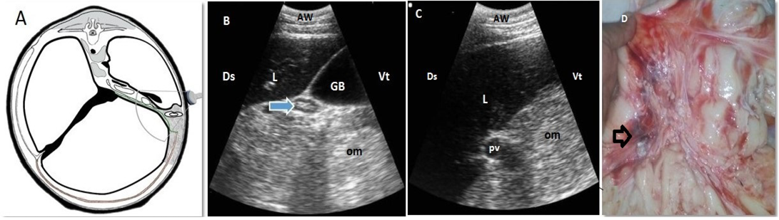
Omental fat necrosis “lipomatosis” of 3-years and 6-months-old-cow. A. Cross-section schematic drawing of abdomen at the level of 13th thoracic vertebra. Note the location of the disease and the transducer. B, C. Ultrasonogram of ventral part of right 10th intercostal space. Note presence of echogenic masses within the omentum (om) with invisible intestinal loops and a compressed duodenum (arrow). D. Lesions presented at necropsy. Note the reddish and blackish discoloured omentum fatty tissues (arrow). AW: abdominal wall; L:liver; GB: Gallbladder; pv: portal vein, Ds: dorsal; Vt: ventral
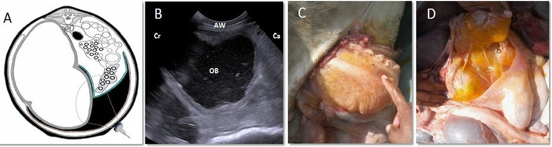
Omental bursitis of 4-years-old-cow. A. Cross-section schematic diagram of abdomen at the level of 4th lumbar vertebra. Note the site of inflamed bursa and the location of transducer. B. Ultrasonogram of the same location showing an extensive collection of anechoic exudate with echogenic debris filling the space between superficial and deep layers of greater omentum. C, D. Lesions noticed at necropsy. Note presence of enlarged sac “omental bursa” containing yellowish, gelatinous exudate. AW: abdominal wall; Cr: cranial; Ca: caudal; OB: omental bursa


