Advances in Animal and Veterinary Sciences
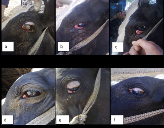
Showing gross appearance of eye carcinoma in (a,b,c,d,e and f) which revealed the shape of carcinic mass after each injection of the ivermectin at a dose 200 mg/Kg continuously with time decrease the tumor mass
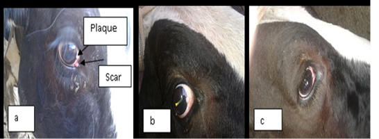
a) represent the lesion in the limbus (sclera-corneal junction) as a plaque and carcinic mass in the third eyelid appear as a scar. (b and c): represent the degradation of the lesion after treatment with two doses of ivermectin result in clearance of the eye.
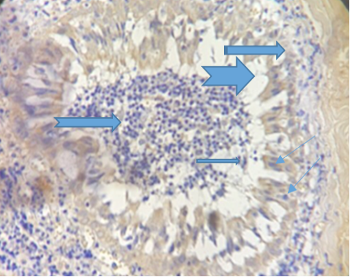
Photogarph for immunohistochemical section of conjunctiva revealed presence of high number of goblet cells ( ) with sloughing of vacuolated epithelial cells () lining conjunctiva in the lumen contain inflammatory exudates () .(Dapi stain X10).
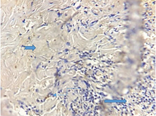
Show congestion of the blood vessels ( ), vaculation () and presence of sebaceous gland (). (Dapi stain X10).
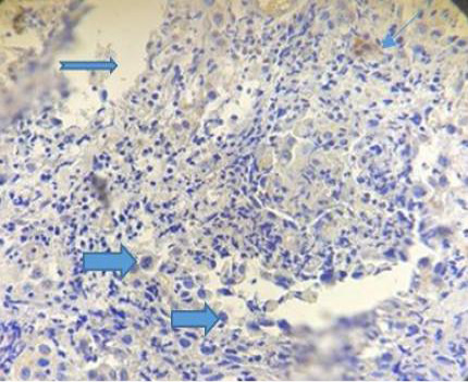
Photograph for immunohistochemical section of conjunctiva showing sever exudation ( ), vascularization () and fibrinous deposition (). (Dapi stain X10).

Photograph for immunohistochemical section of conjunctiva showing sever exudation ( ), vascularization () and fibrinous deposition (). (Dapi stain X10).
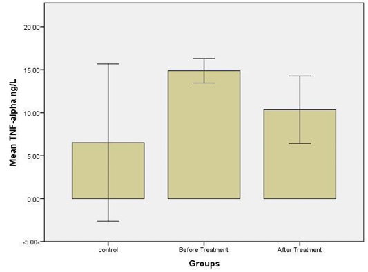
Revealed Levels of TNF-Alpha. Significant differences at (P≤0.05).


