Advances in Animal and Veterinary Sciences
Research Article
Adv. Anim. Vet. Sci. 7(5): 378-382
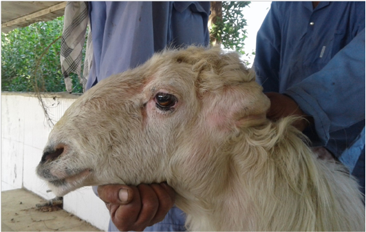
Figure 1
Photo of Awasi ram infected with Pseudomonas aeruginosa showing marked enlargement of retropharyngeal lymph node.

Figure 2
Cross-section of retropharyngeal lymph node of Awasi ram infected with Pseudomonas aeruginosa showing abscesses in the medulla and cortex of the node.

Figure 3
Photomicrograph of the affected lymph nodes of Awasi sheep infected with Pseudomonas aeruginosa showing: area of suppuration of lymphoid tissue (A), medullary area in which the sinusoids were rich in neutrophils and macrophages and severe congestion and edema (B), suppuration of lymphoid tissue, granulomatous inflammatory reaction and congestion (C), anddilated sinusoid (due to edema) in medulla with some macrophages and neutrophils (D) (H&E stain 200X).


