Advances in Animal and Veterinary Sciences
Research Article
Adv. Anim. Vet. Sci. 7(2): 106-111
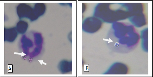
Figure 1
Anaplasma phagocytophilia organisms in a neutrophil (A) and Monocyte (B) Buffy coat smear; Giemsa stain. Magnification, 1,000.
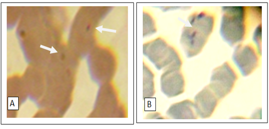
Figure 2
Numerous small basophilic structures of hemotropic Mycoplasma spp. organisms on the surface of the erythrocytes attached to cell membranes singularly(A) or in clustered (B). Peripheral blood smear; Giemsa stain. Magnification, 1,000.
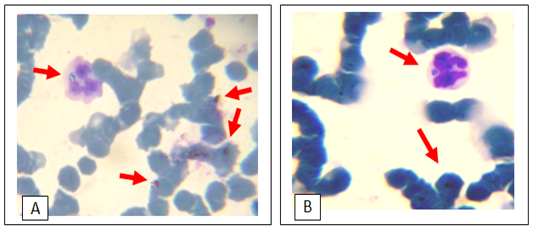
Figure 3
Giemsa stain light micrograph (Magnification, 1,000) of horse blood smear, co-infection of hemotropic Mycoplasma spp. and A. phagocytophila
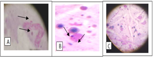
Figure 4
Giemsa stain of tick hemolymph cells infected with Anaplasma phagocytophilium (A), hemotropic Mycoplasma spp. (B) and co- infection of both bacteria (C)


