Advances in Animal and Veterinary Sciences
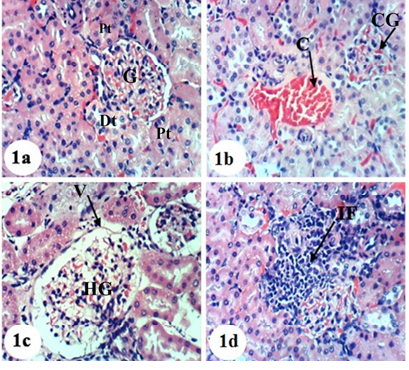
Photomicrographs of H&E stained kidney sections of normal and APAP-administered rats. 1a: Photomicrograph of kidney section of normal rats showing normal histologic structure of kidney; normal glomerulus (G), normal proximal tubules (PT) and distal tubules (DT) 1b-d: Photomicrograph of kidney section of APAP-administered rats showing congestion of renal blood vessel (C), congestion of glomerulus (CG) in b, hypertrophied glomerulus (HG), vacuolization of the endothelial cells lining the glomerular tuft (V) in c and interstitial nephritis (IF) in d. ×400
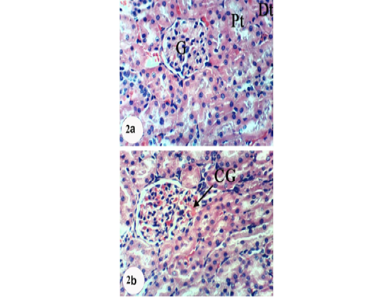
Photomicrographs of H&E stained kidney sections of APAP-administered rats treated with navel orange peel hydroethanolic extract. 2a: Photomicrograph of kidney section showing slight congestion of glomerulus (CG). 2b: Photomicrograph of kidney section showing normal structure of kidney; normal glomerulus (G), proximal (Pt) and distal tubules (Dt). X400

Photomicrographs of H&E stained kidney sections of APAP-administered rats treated with naringin. 3a: Photomicrograph of kidney section showing congestion of renal blood vessel (C) and congestion of glomerulus (CG). 3b: Photomicrograph of kidney section showing normal structure of kidney; normal glomerulus (G), proximal (Pt) and distal tubules (Dt). ×400
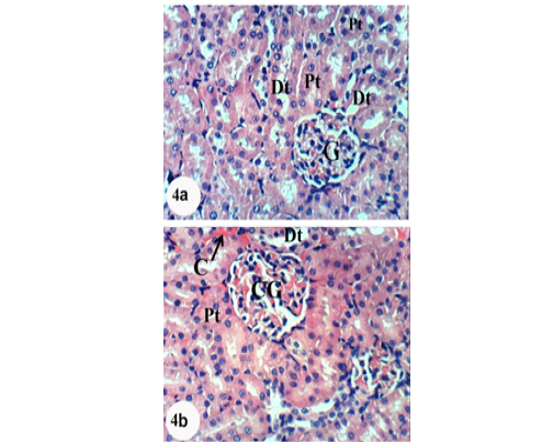
Photomicrographs of H&E stained kidney sections of APAP-administered rats treated with naringenin showing normal structure of kidney; normal glomerulus, proximal and distal tubules. Slight glomerular and intertubular congestion was observed in 4b. X400


